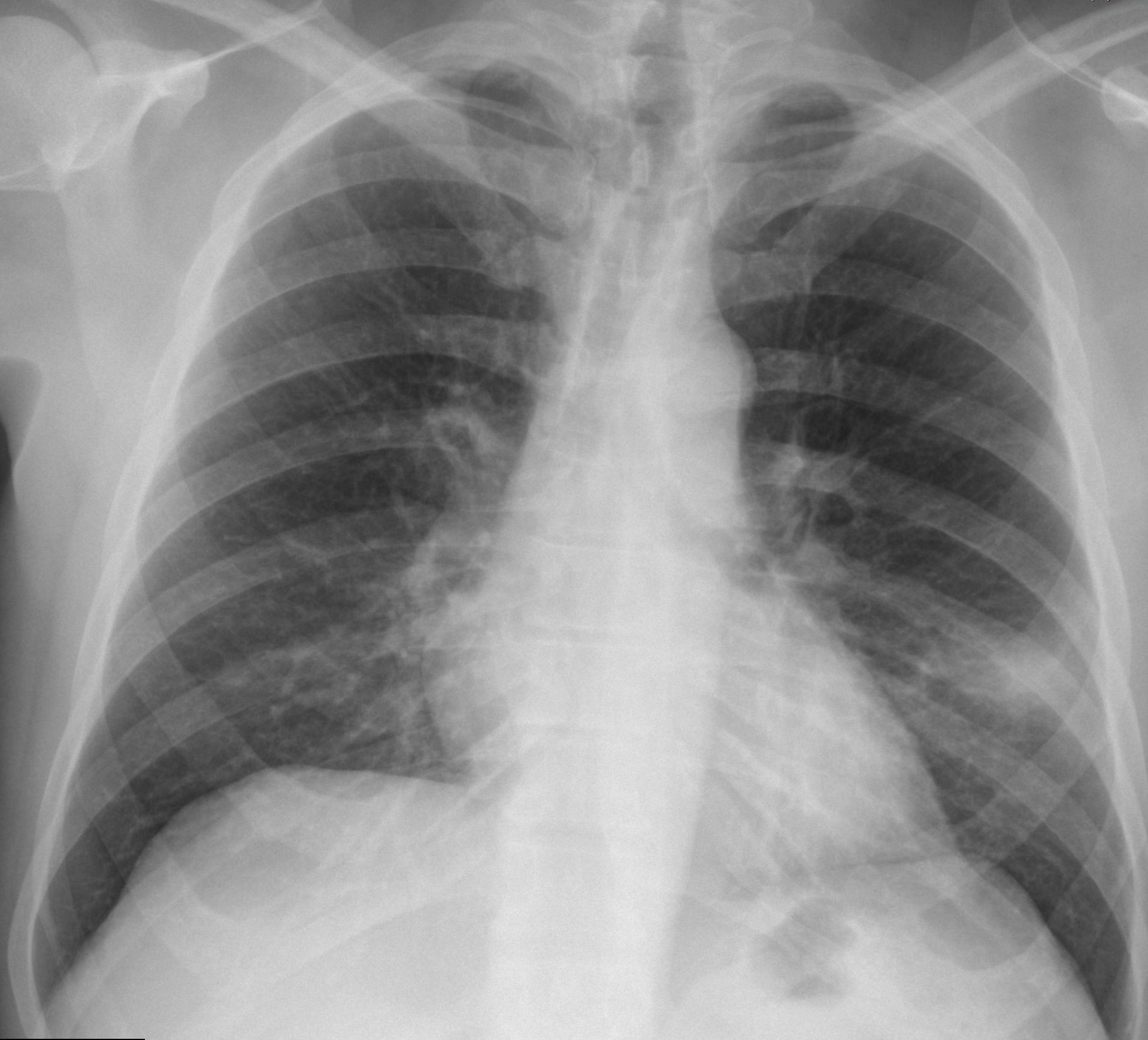
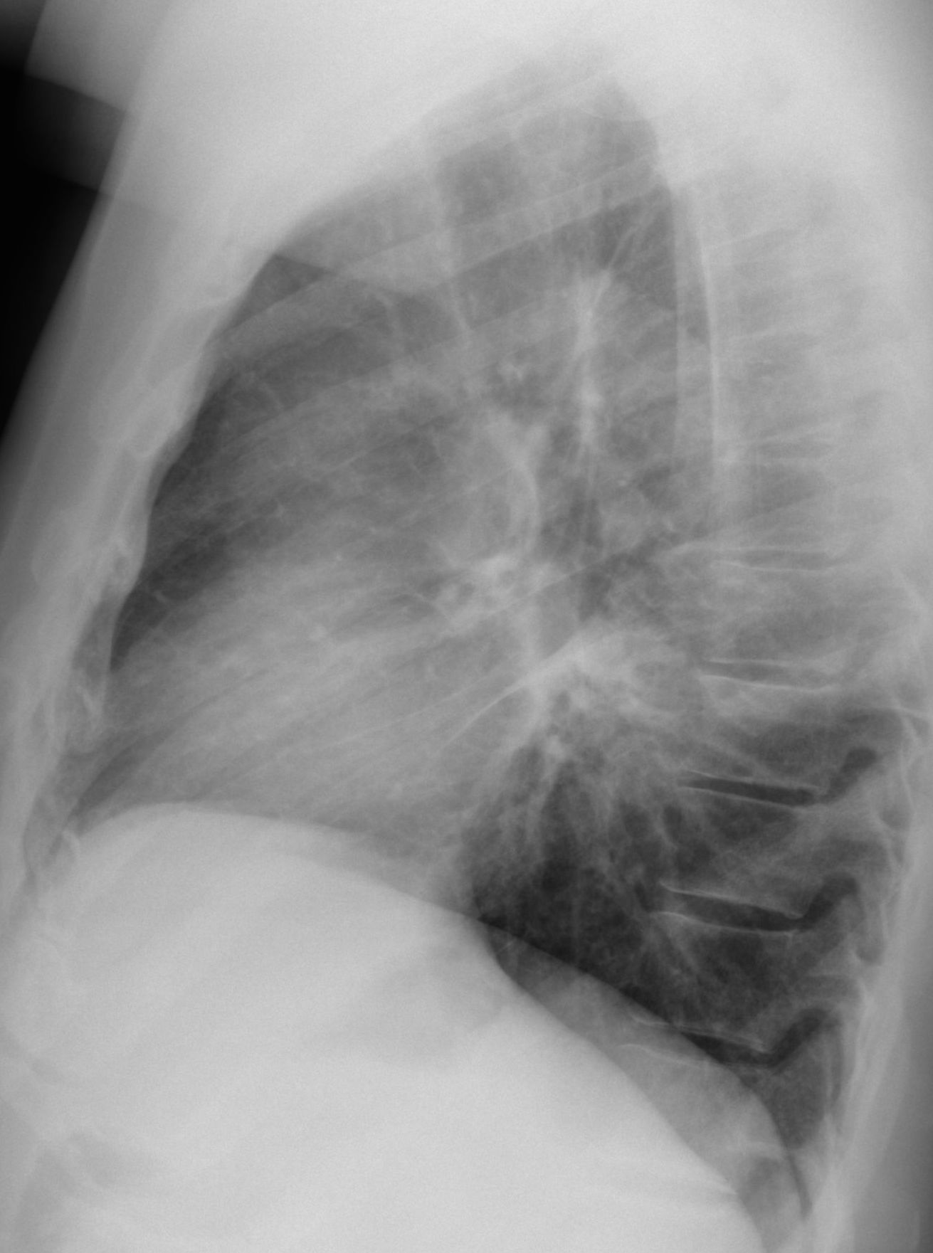
Muppet is very excited because case 100 is only two weeks away. In the meantime, he wants to present a simple case that we saw one month ago. The radiographs below belong to a 53-year-old man with cough and fever. What do you see?
Leave your thoughts in the comments and come back on Friday for the answer.
Findings: PA radiograph show a segmental air-space opacity (A, arrow) which, given the clinical information, represents most likely a pneumonia. In addition there is buckling of the upper trachea (A, red arrow). The cone down view shows the tracheal angulation and a rounded air-filled structure at this level (B, arrows).
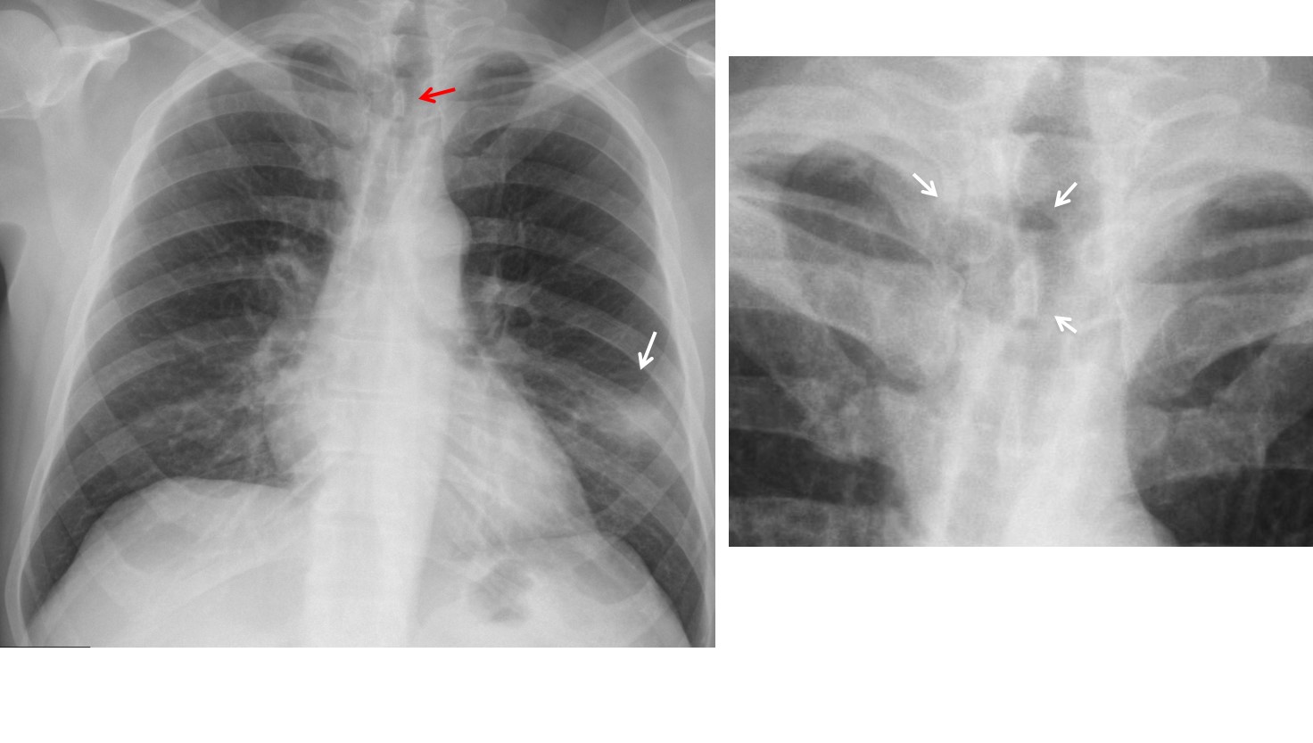
Answer – A & B
Axial and coronal CT confirm the presence of a diverticulum impinging on the trachea (C, D arrows).

Answer – C & D
Tracheal diverticula are uncommon and incidental findings, found in less than 1% of patients at autopsy. Usually asymptomatic, occasionally they accumulate secretions that may became infected.
Final diagnosis: Segmental LLL pneumonia and tracheal diverticulum
Congratulations to Genchi Bari, who made a good diagnosis
Teaching point: always look at the trachea in the PA and lateral films. You may discover interesting pathology.