Dear Friends,
Muppet is very happy with your progress. But, given his mean disposition, he wants to test you with this case:
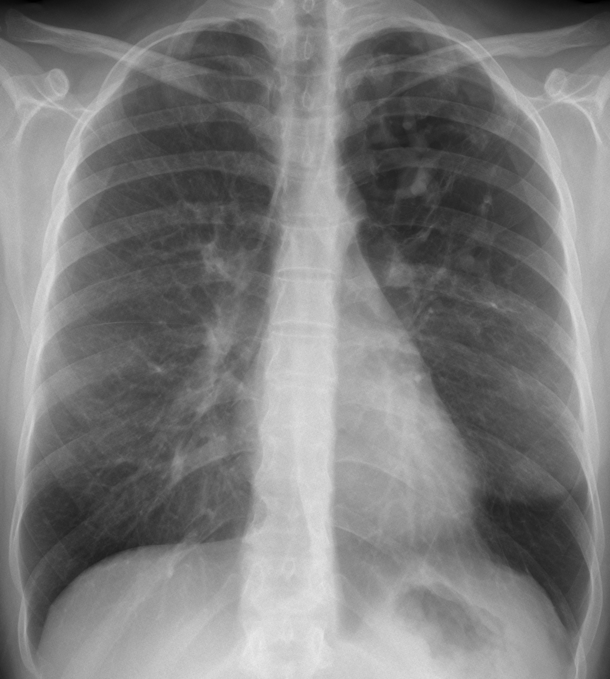
Forty-year-old woman with a moderate cough.
Diagnosis:
1. Tuberculosis
2. Vascular malformation
3. Aspergillosis
4. None of the above
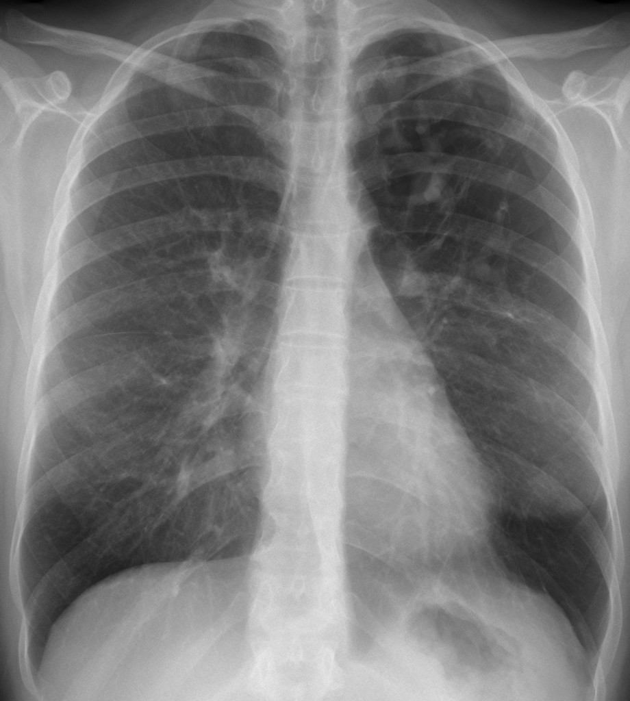
40-year-old woman, PA chest
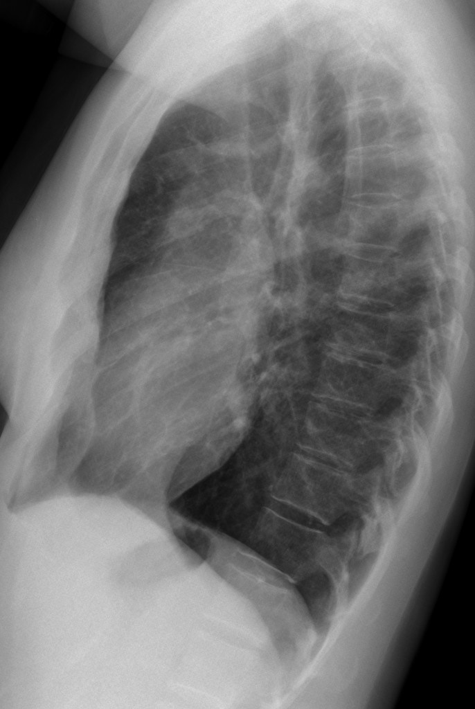
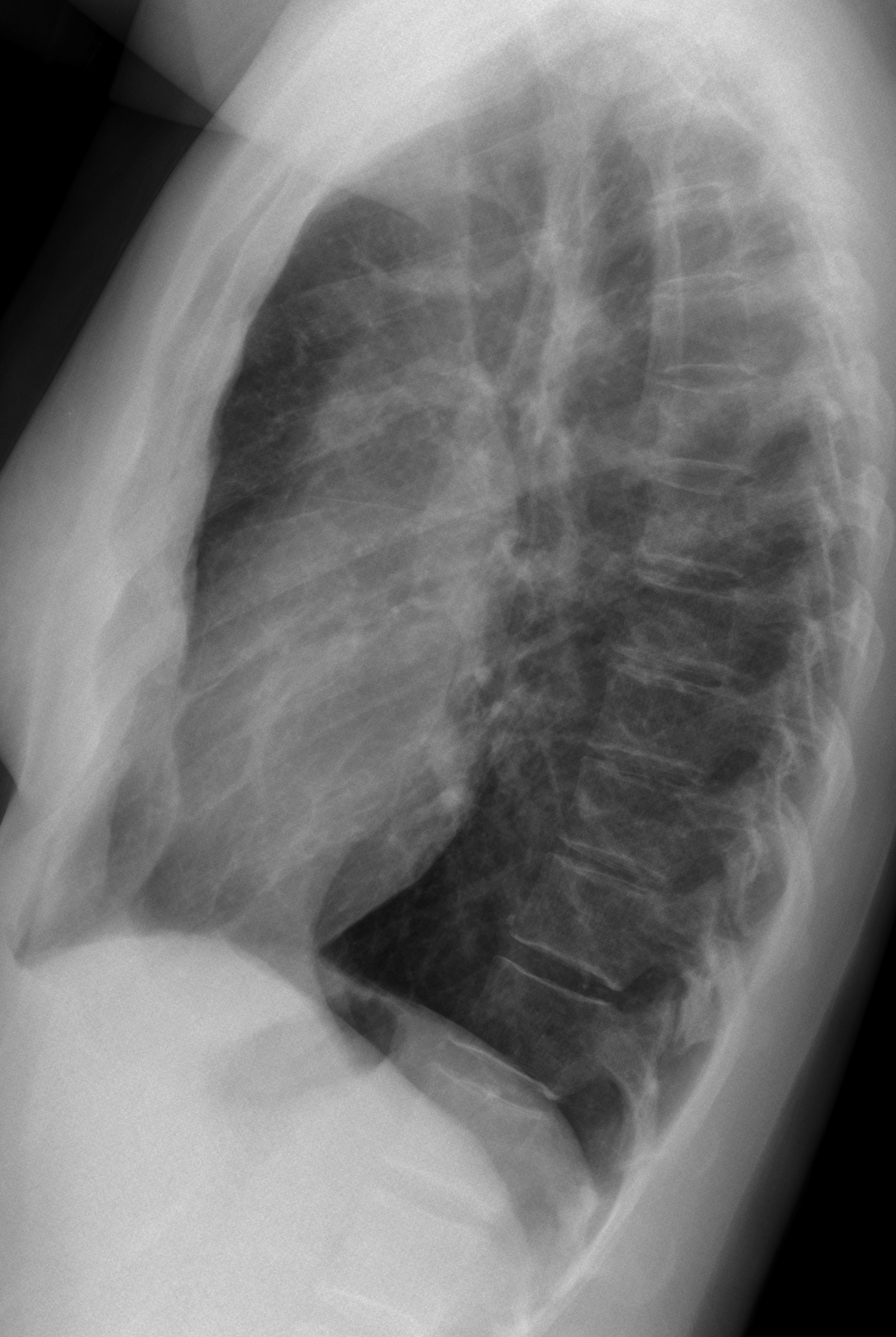
40-year-old woman, lateral chest
Click here for the answer to case #13
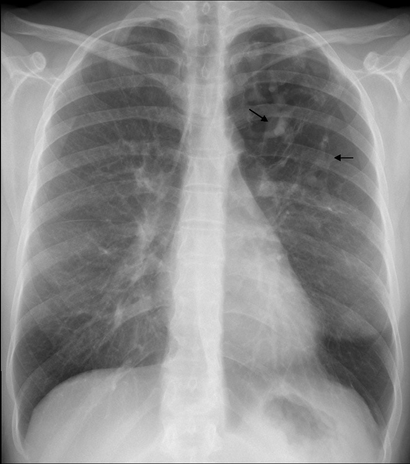
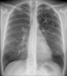
The PA chest film (fig. 1) shows an area of hyperlucency in the left upper lung, with branching, serpiginous shadows within (arrows). Whenever we see branching shadows, two possibilities should come to mind: vessels or dilated bronchi. Vascular malformations are not associated with hyperlucent lung, whereas the combination of branching shadows and hyperlucency should make segmental bronchial obstruction with mucous impaction the most likely possibility.
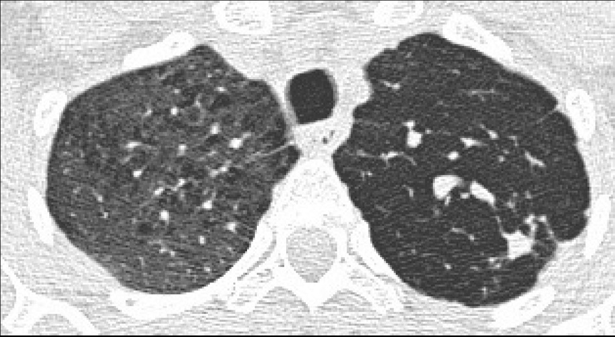
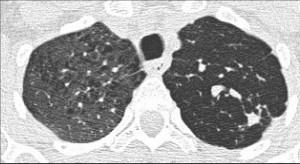
Axial and coronal enhanced CT confirms the unenhanced mucous plug (figs. 2,3 arrows) within the area of hyperlucency. Expiratory film confirms air trapping (fig. 4). Final diagnosis: congenital bronchial atresia, confirmed with bronchoscopy.
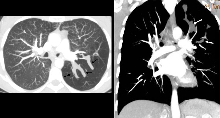
Teaching point: think of mucous impaction when you see branching shadows.
Remember that the most common cause of mucous impaction is lung carcinoma. When in doubt, perform bronchoscopy.