Dear Friends,
Sorry about the delay in posting a new case, but Muppet was invited to the ECR, got drunk every night and did not cooperate at all. By the way, he sends his warmest regards to Marina, a very smart resident.
Our new patient is a 36 year-old lady with lupus, admitted with mild dyspnea.
Diagnosis:
1. Myocardiopathy
2. Pericarditis
3. Hilar adenopathy
4. None of the above
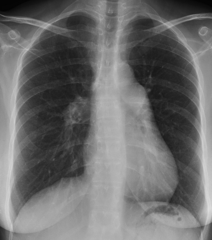
36 year-old woman. PA chest
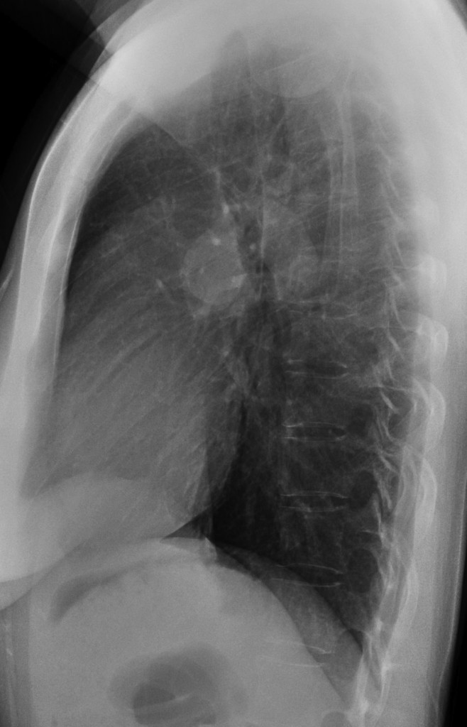
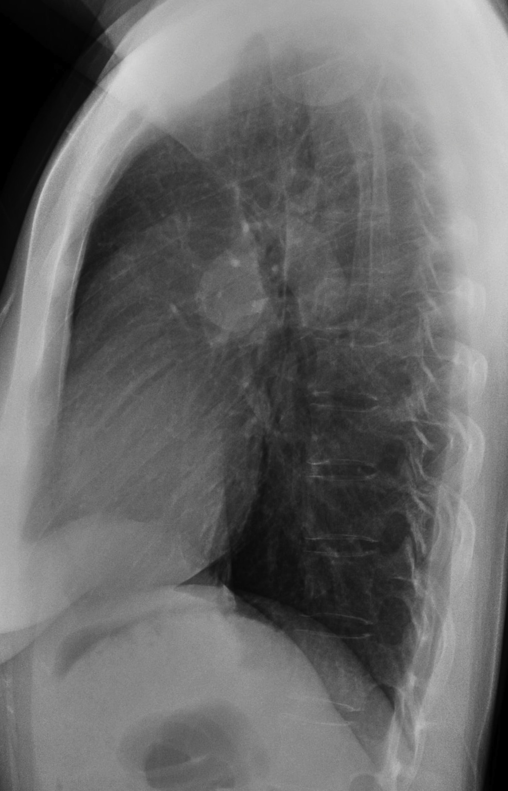
36 year-old woman, lateral chest
Click here for the answer to case #14
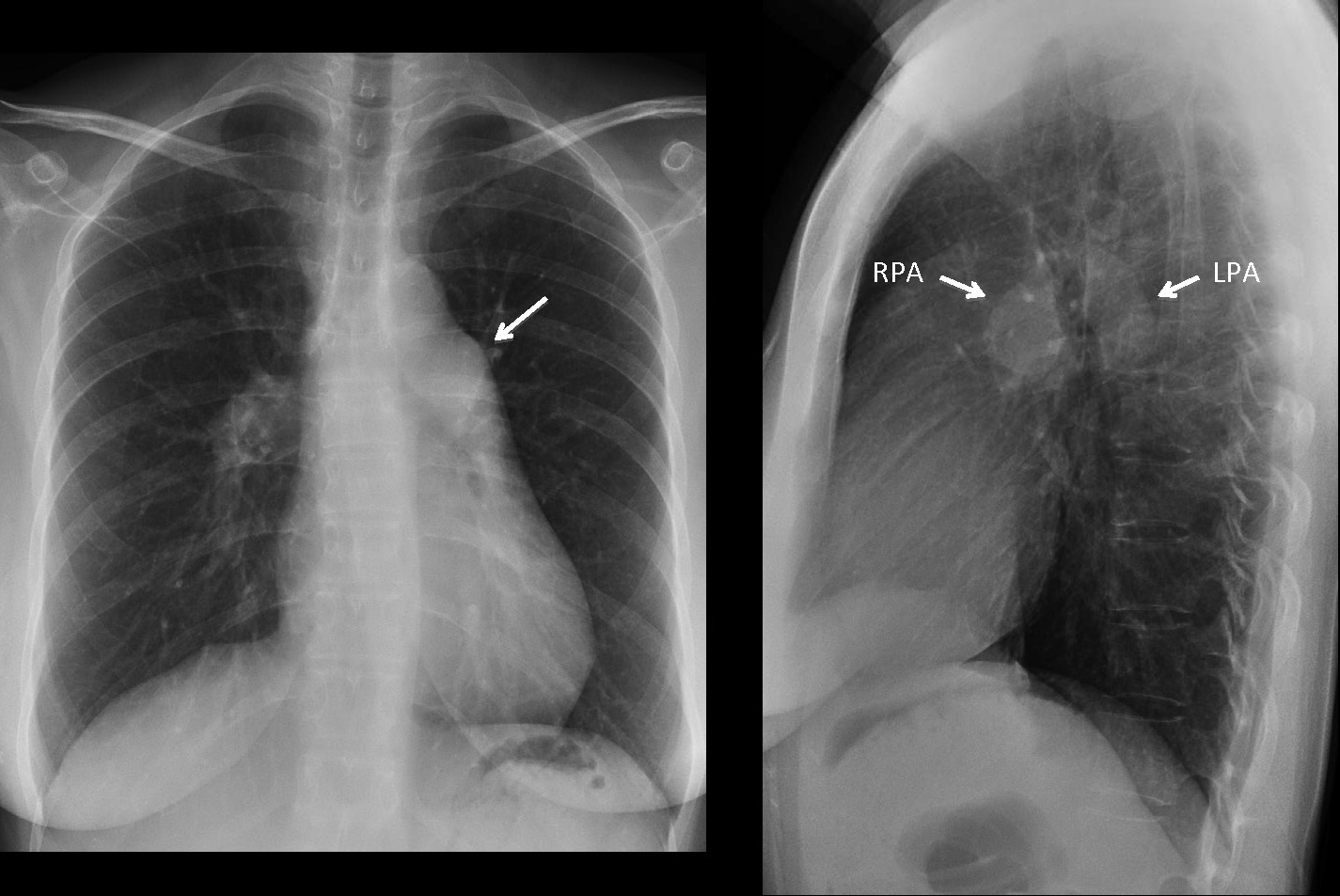
The PA chest radiograph shows convexity of the pulmonary arch (Fig 1 arrow), as well as prominent central pulmonary arteries, with diminished vascularity of the lungs.
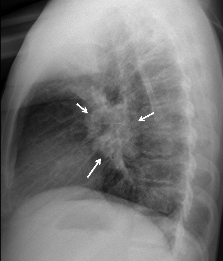
This appearance is very typical of pulmonary arterial hypertension. The lateral view is very helpful because it helps to differentiate between the enlarged right and left pulmonary arteries (Fig 2 arrows) and lymph nodes, which present as the ‘donut sign’ (Fig 3).
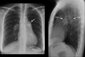
Fig. 1&2
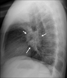
Fig. 3
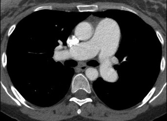
Enhanced CT (Fig. 4) confirms the increased size of the main pulmonary artery, which measures 34.5mm in diameter.
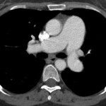
Fig. 4
Pulmonary arterial hypertension has many causes, among them COPD, chronic pulmonary embolism, idiopathic, inverted shunt, vasculitis, etc. In this particular patient, the pulmonary hypertension was ascribed to lupus vasculitis.
Teaching point: to differentiate PAH from enlarged lymph nodes a) look at the mediastinum to see if other lymph nodes are present, and b) look for the ‘donut sign’ on the lateral view, which is a sign of enlarged lymph nodes.