Dear Friends,
Muppet is getting soft in his old age and presents you with an easy case: pre-op chest in a 40-year-old male with a brain tumour.
Diagnosis:
1. Lung metastases
2. Lung carcinoma
3. Pleural tumour
4. None of the above
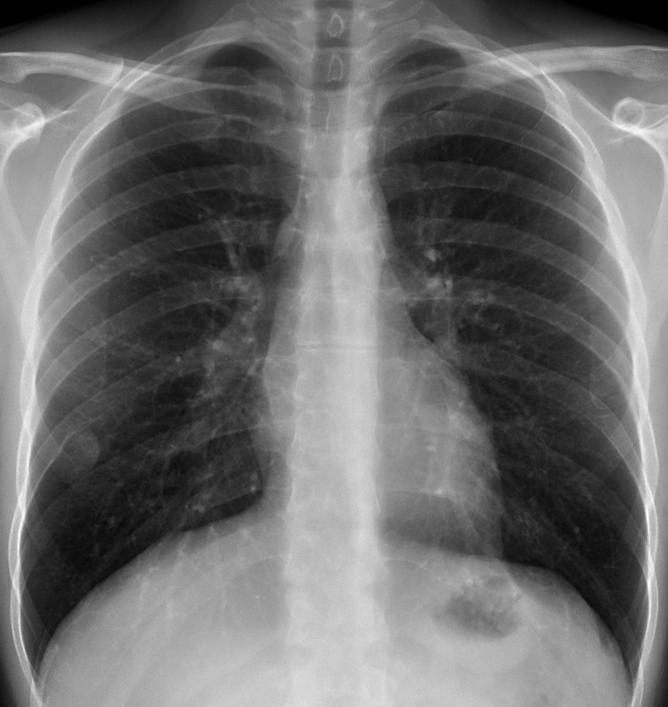
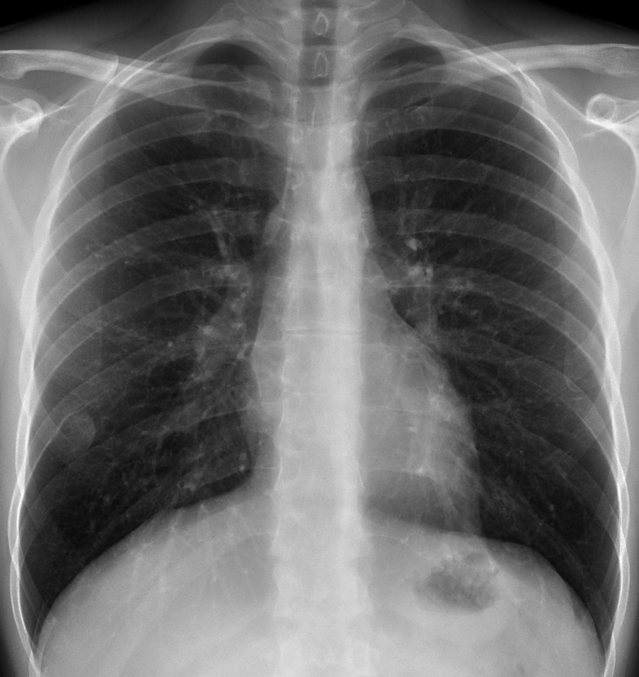
40 year-old man, PA chest
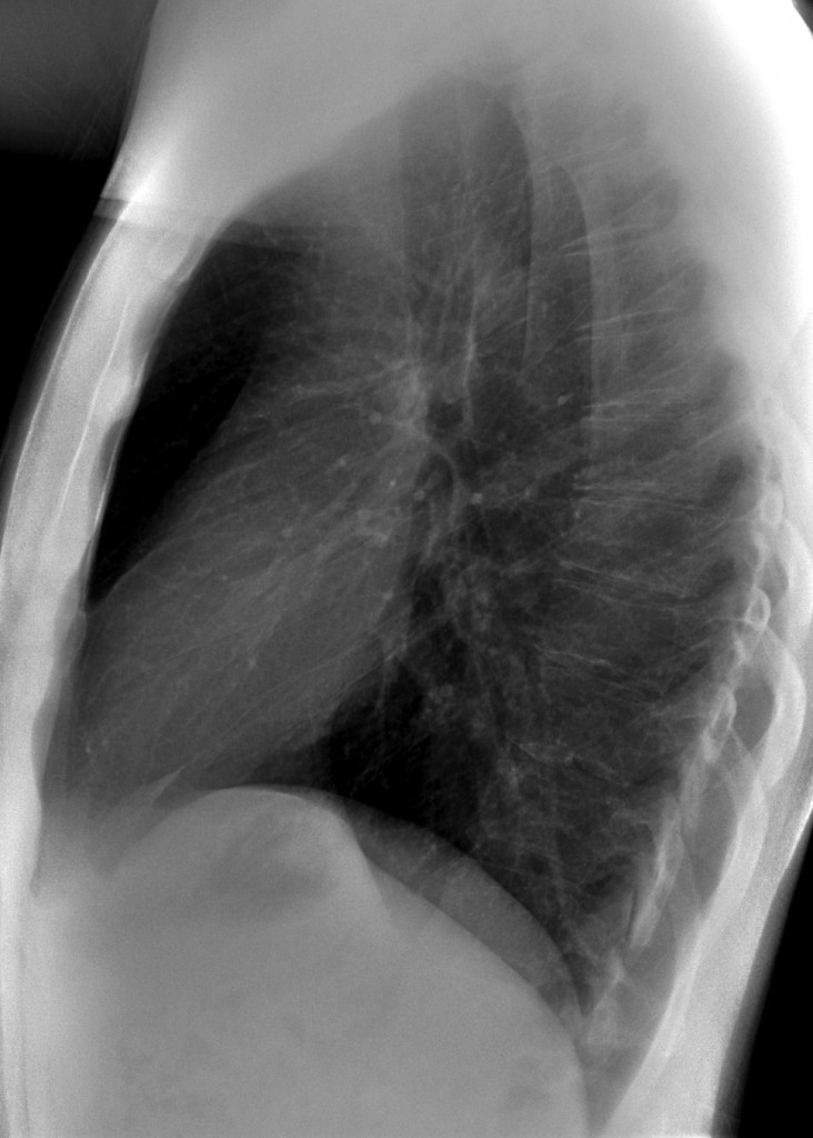
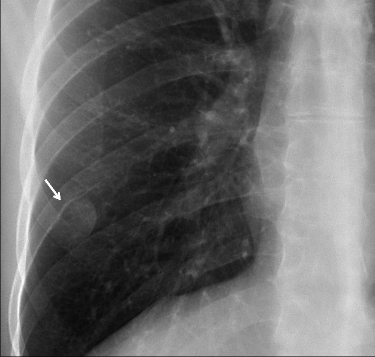
40 year-old man, lateral chest
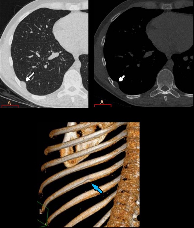
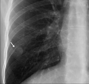
The PA chest film shows a rounded lesion with the ‘incomplete border sign’ (medial aspect outlined by air, lateral border not visible because in contact with chest wall). In addition, there is erosion of the lower border of the rib (arrow). This combination of signs is pathognomonic of a mass in the underside of the rib. CT confirms the presence of a soft-tissue mass and the erosion of the rib (arrows).
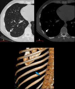
Final diagnosis: neurofibroma. The patient had neurofibromatosis and a meningioma. Muppet is very happy with your diagnostic skills and your semiological interpretation.
Teaching point: when the underside of the rib is eroded, the lesion arises from a structure which travels under the rib: a nerve (as in this case), vein, or artery
(remember the erosion in aortic coarctation).