Muppet gratefully acknowledges the contribution of Dr. Jose Vizuete and Dr. Vilar, who supplied this case (Muppet also wants to point out that any complaints should be addressed to them!).
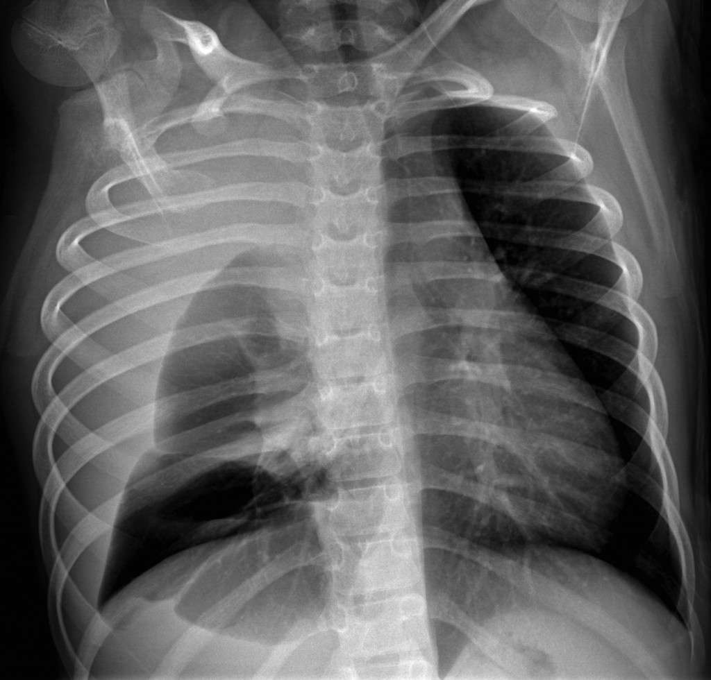
AP chest of an 11-year-old girl with dyspnea for the last week and mild fever.
1. Staphylococcal pneumonia
2. Mediastinal mass
3. Foreign body
4. None of the above
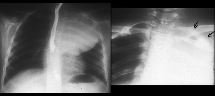
The AP chest radiograph shows partial opacification of the right hemithorax, with probable pleural effusion. The trachea and mediastinum are displaced towards the left, indicating a probable mass in the right hemithorax. The second rib is abnormal (arrows), suggesting that the mass arises from the chest wall. Findings are confirmed with CT, which shows a huge mass and the alteration of the rib (arrows).


In a child, the combination of a large mass with affectation of a rib is suggestive of Ewing’s tumour or Askin tumour. Surgical diagnosis: Askin tumour.
In his (lost) youth, Muppet saw two similar cases of Askin tumour, both with large masses and not very obvious rib involvement (Fig. 3).

Fig 3. Askin tumour presenting as a large mass and affectation of the first left rib (arrows)
There is a similar case in the paper ‘Pediatric ribs: a spectrum of abnormalities’ (RadioGraphics Jan 2002 22:887-104). In his limited experience, Muppet suggests that if you see a child with a large tumour and limited affectation of a rib, think of Askin.
Before anybody starts complaining (case too difficult, I don´t see the abnormality, it’s easy to see the findings after looking at the CT, etc.) Muppet would like to point out that Dr. Vizuete saw the findings in the plain film and suggested the diagnosis.
Teaching point: when you see large masses that contact the chest wall, look at the ribs. They may give you a clue.


AP, but is it made in dorsal decubitus or in ortostatism?
Mediastinal mass, because I can see the trachea deviated towards the left (and maybe slightly posterior) – deviation too big to big caused by the amount of fluid in the hydro-penumothorax.
i think non of the above
I think it may be a right foreign body
there is tracheal deviation
but if it was mass or fb aspiration why the lung is not toally collapsed
why it is not white lung ?
i need the answer doc 😀
Be patient. While you wait, look at the ribs.
This case is more difficult..!! Could be a mediastinum mass who makes dislocation of trachea, a lot of pleural effusion and hernia of right chest..
there is also a very big right hilum
i can’t look the liver area and the gastric bulla (hernia??) and instead, is important if this is ortostatic or not
4. None of the above
The trachea and mediastinal structures are displaced to the left, the right lung is colapsed and I think is due to a tension pneumothorax,furthermore the pleural cavity is occupied by pleural efusion. But, because does not adopt the air-fluid level, I think this RX could be done in decubitus??.
looks like left diaphragmatic hernia
rt diaphragmatic hernia
The position of the arms suggests a standing position. There are at least 2 air-containing right lobes, probably 3. No infiltration or cavitation is seen. The amount of free-flowing pleural fluid is relatively small. The skeleton is normal, but there is probably some scoliosis and torsion in the spine. No signs of air in the pleura. There must be large, space-occupying solid or encapsulated and at least partly well-demarcated pleural mass. My answer is: None of the above.
Very interesting
I dont think its a collapse due to forign body coz left ling is still can be seen, dont think its pneumonia.
Mediastinal mass is a possibility however i cant explain the big opacity on the right side peripherally unless the mass is coming from posterior mediastinum & pushing the whole right lung forward.
A lateral view x ray would be very helpful
none of da above lateral view wud b usefull to give diagnosis…
Idro-pneumotorace ipertensivo, da fistola bronchiale (TBC ?)
My head is revolving! I am seeing a left cervical rib.
Coming to the image on a more serious note- It is not the pneumatocele of staph pneumonia , nor is it a foreign body associated collapse.Mediastinal mass- I am not seeing mediastinal widening, it is shift of mediastinum to the left.
The point that increases my curiosity is that if there is a pleural collection, then the underlying lung should be collapsed atleast partially. I have never seen transluscent shadow like this in a collapsed lung. So there is definitely some air collection ?posteriorly or in the the lung itself. A hernia will come from down but the air collection seems tob be bounded inferiorly.
Again one more thing that is making me curious is that my eyes are seeing something which I am forced to believe are the fissures on right side. (unless they are fibrotic strands at the exact locations of the 2 fissures!)
The answer seems to be none of the above but what then is it?
Of course I will continue to think of any syndrome of the childhood/ some infective condition(not sure if the infection is primary or secondary in the infected lungs)….
Right lung hypoplasia ???
On 2nd thoughts, the anterior ends of 2st and 2nd ribs are broad when compared to their left counterpart. The thing that still puzzles me is why on the 1st place was an ap film ordered? Am trying to find the reason for that. Maybe the clue to the diagnosis lies somewhere in ordering the ap film.
Again the opacity does not seem to be pleural and the underlying lung if any may be hyperinflated or air collection in some form may be present in there.
I am not able to trace the right scapula other then its acromion process!
In the upper part of luscent shadow i can see some soft rounded opacity on the right side.
1st rib on the right seems to be abnormal…overlapping of 1st rib with scapula does not seem to be normal.The coracoid process…or is it a bifid 1st rib or my inexperience in reading ap films!
Still thinking…
Keep on thinking. You are getting closer…
AP or PA do not influence the findings Muppet says
Trying to locate the right clavicle and medial ends of both 1st ribs….
Still thinking..
See below…
osteogenesis imperfecta
I hope you don’t consider this a easy case to diagnose on a xray, i don’t…
AP projection, decubitus, chest slightly rotated to the right.
Large right pleural effusion. Complete atelectasis of middle lobe, maybe partial also RUL. Lucency of the right bronchia not visible, obscured by a right paratracheal +right hillar mass (probably lobulated ADP complex) displacing the mediastinum to the left; since this is a child, thymus must also be considered, although it should not displace trachea so far; angle of the carina is reduced, further indicating large paratracheal mass. Transudative right pleural effusion could occur in the compression of the right brachiocephalic vein (is there also edema of the arm?)
Poorly contoured predominantly expansile lucent lesion of the antero-lateral arch of the 2nd right rib with irregularity of the bone contours at the anterior margin but no periostal reaction. At first, the rib lesion seams to me a monostic fibrous dysplasia; dx of bone lesion: 1) since there is also fever, the possibility of osteomyelitis cannot be excluded (although the lack of peristeal reaction would say otherwise) 2) Invasion of a anterior mediastinal mass ??
So no clear diagnosis from me, i think though there is a high probability of lymphoma, but that usually gives the ivory aspect on the ribs / also it could fit a thymic carcinoma but that rarely occurs in children. Very intriguing findings (partly because i can’t see the big picture), i’m curious to see what it was on CT…
Well done! See you have overcome your SOS syndrome!
What’s the relationship of rib lesion with mass? Lymphoma is a possibility, but there are others.
OK. Waiting for muppet’s answer..It sure is a very interesting case. A lateral view would have been helpful and also an apicogram. But then these are just wishes….
There seems to be a mass at T3, T4 level on the right pushing the trachea. ?Thymoma ?Lymphoma ?retrosternal thyroid
None of the above is my answer.
Could be a right diaphragmatic hernia?
rib involvement – shift of mediastinum – malignancy?
I think there is a high mediastinum mass.
Then, is it possible that all the right chest wall was like distended?
I’m thinking in a mediastinal mass that’s occupying also the right EXTRApleural space (I cannot believe there is pleural effusion or chylothorax, or any occupation of the pleural space because I think there must be some collapse of the lung; not the same for extrapleural occupation).
It reminds me a kind of exaggerated “signo de la ola tímica” -can’t translate- and I would think of thymoma.
Did not see the rib…sorry.
I think the bone affection is the typical described in small cell round tumors (Lymphoma and PNET (including Askin Tumor- Ewing) wich should show extensive soft-tissue and bone expansion without or with minimal cortical destruction.
I keep on with my idea of the extrapleural mass.
I’m not sure of anything at all…
I think there are a large right pleural effusion, right lung compression, right paratracheal and right hillar mass, dislocation of mediastinum to the left. And what about the 10 rib ir the right(posterior arch)? Is it destruction?
leftward displacement of the heart could be secondary to a pectus excavatum.
-massive hydropneumo/pleural effusion with bone lesions may be caused by cystic angiomatosis/lymphangioleiomyomatosis.
maybe pulmonal arterry’s embolia?
Hi,
We had a similar askins case discussed in our local forum.
Great case.
Thank you for the information. Supports the opinion about the binome: large mass, scarce rib affectation
click to view gucci handbag outlet with low price
I’m sure the best for you cheap louis vuitton for more