Dear Friends,
Muppet is in a good mood today (Miss Piggy may be responsible for that) and wants to present a ‘wysiwyg’ case (what you see is what you get).
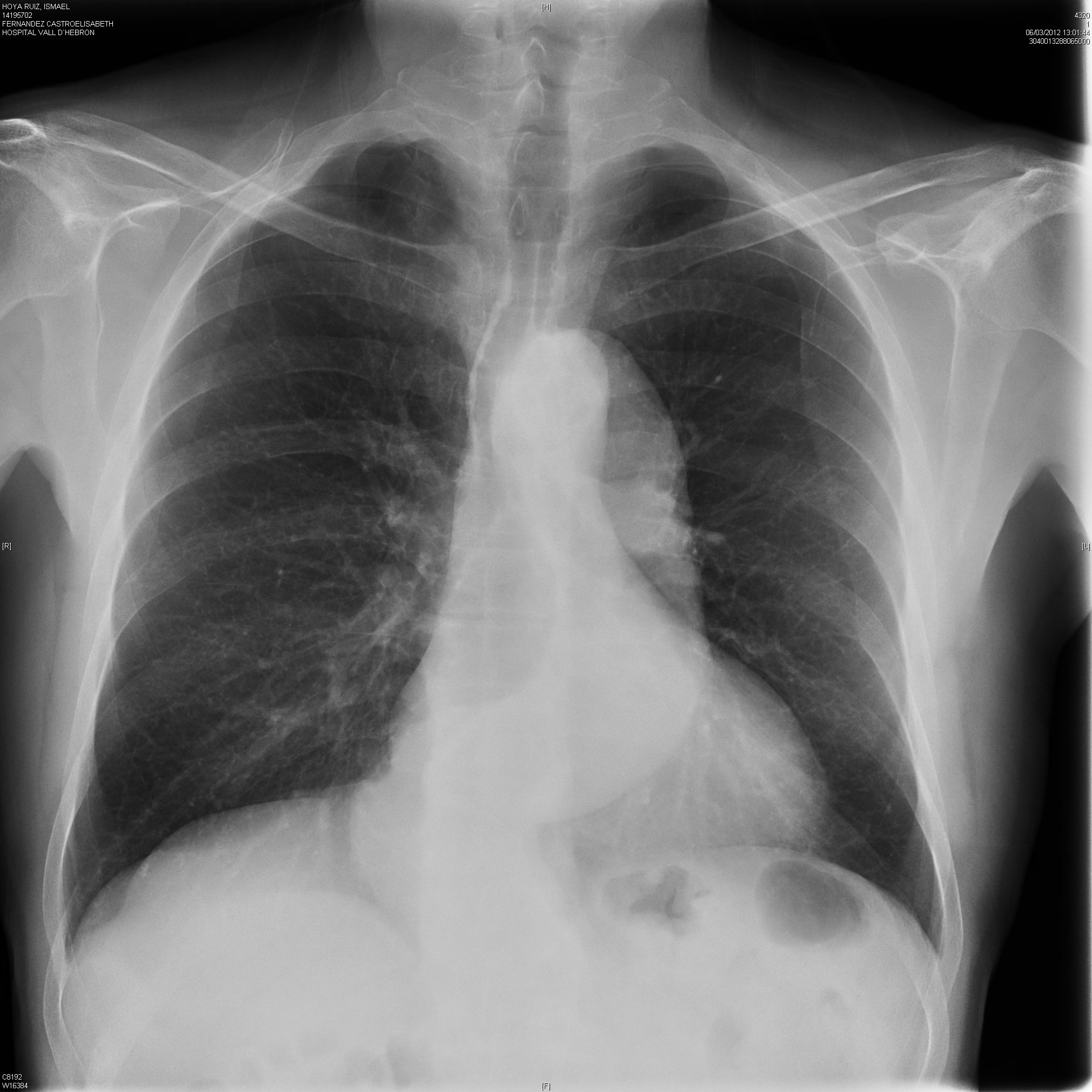
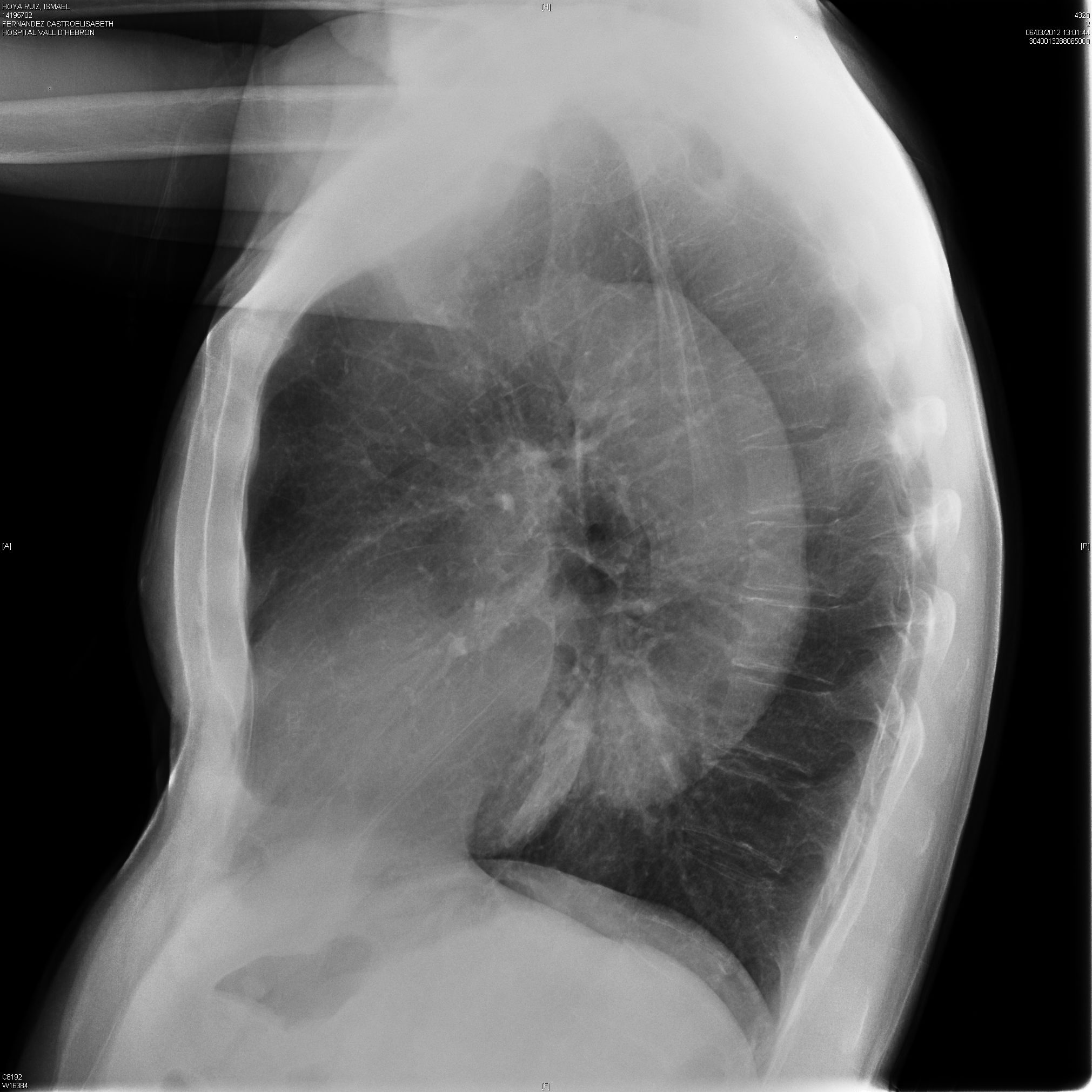
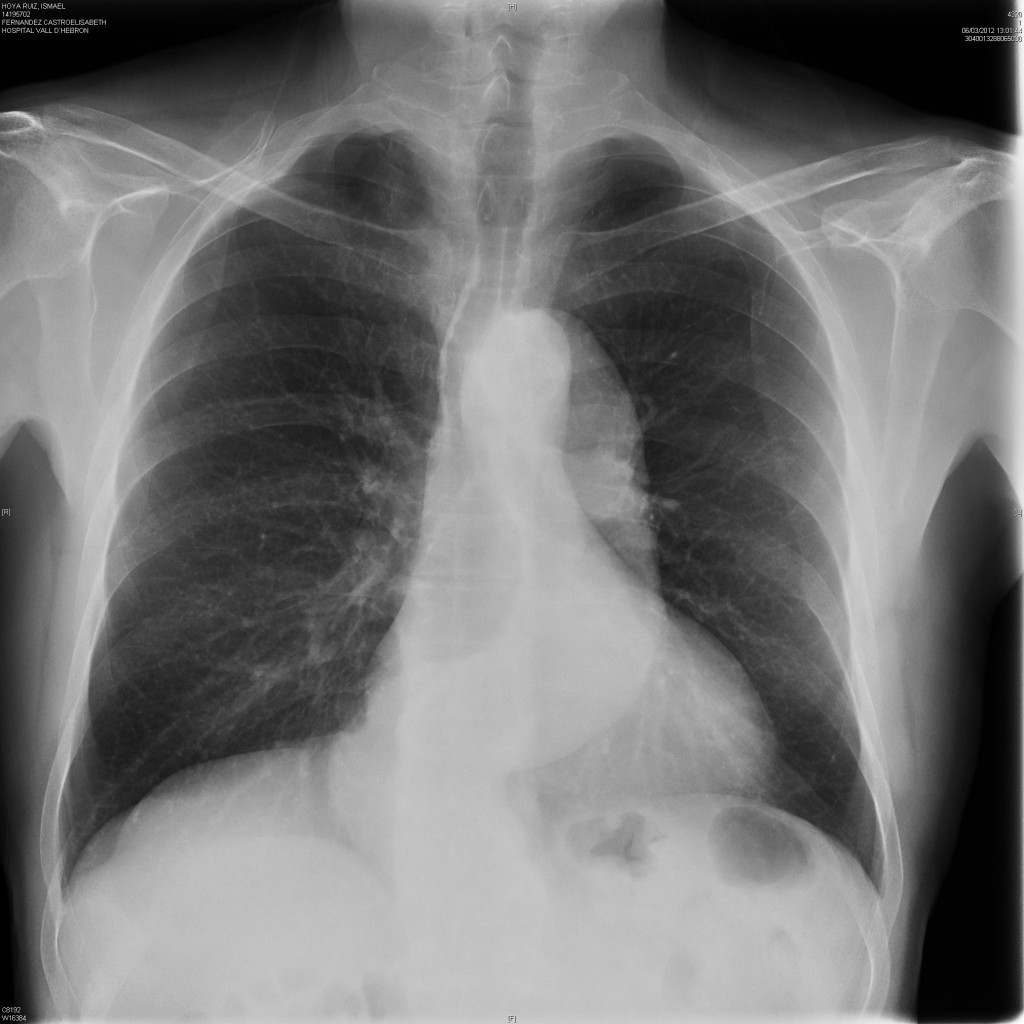
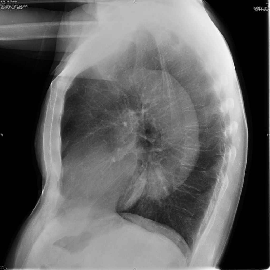
64-year-old, asymptomatic male. Chest images taken one month after an episode of myocardial infarction.
Diagnosis:
1. Aortic aneurysm
2. Aortic elongation
3. Type A aortic dissection
4. Type B aortic dissection
Clicke here for the answer to case #18
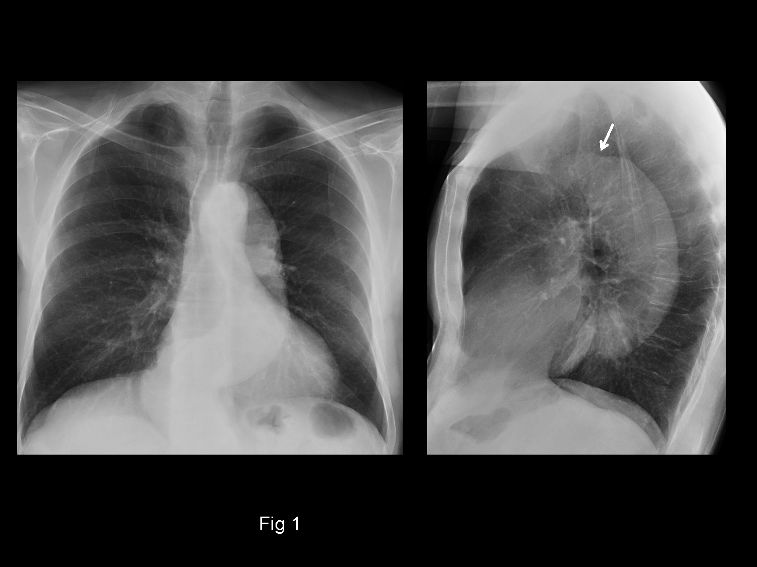
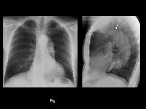
PA and lateral chest show an elongated descending aorta, while the ascending aorta is not prominent at all, as evidenced in both views.
A non-dilated ascending aorta goes against type A dissection and aortic elongation (whole aorta should be prominent). This leaves two possibilities: type B dissection and aneurysm of the descending aorta. In Muppet’s experience, the combination of a non-dilated ascending aorta and dilated descending aorta is highly suggestive of type B dissection. The transition from normal to dilated aorta can be seen in the lateral view (arrow).
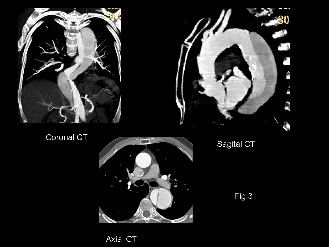
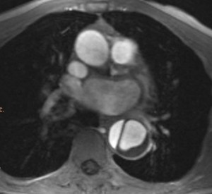
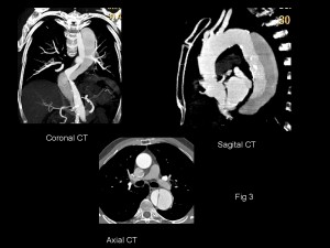
In this patient the dissection was discovered accidentally in a cardiac MRI performed to evaluate the heart after the myocardial infarction (fig 2). Enhanced CT confirms the diagnosis of type B dissection (fig 3).
Congratulations to all of you that made the correct diagnosis, except if you are supporters of Chelsea FC. Muppet is a Barcelona fan and hates losing!
Teaching point: enlarged descending aorta with normal-looking ascending aorta strongly suggests type B dissection, even though the patient is asymptomatic. Enhanced CT should be performed to investigate this possibility.
Also, never play a football match with a defence of three!