Dear Friends,
Muppet wants to test your diagnostic skills with the following case:
75-year-old male with vague pain in the right hemithorax.
Diagnosis:
1. Myeloma
2. Hydatid cyst
3. Fibrous dysplasia
4. None of the above
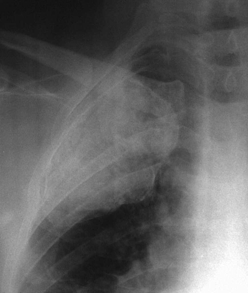
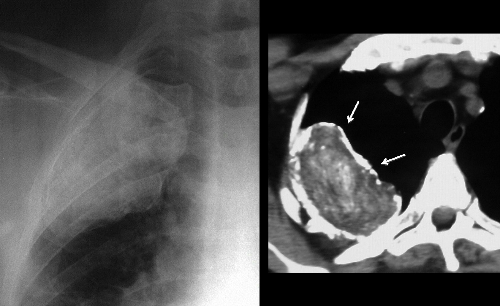
75-year-old male, PA chest
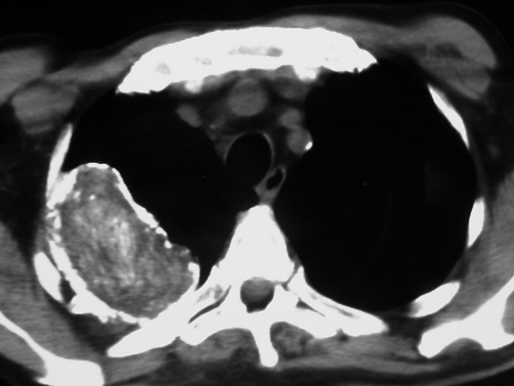
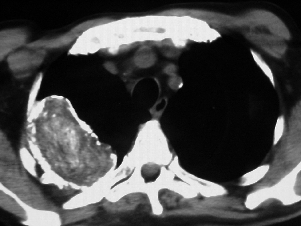
75-year-old male, CT
Click here for the answer to case #19
The PA chest film shows an irregular mass with calcified borders in the upper lung field. The posterior arch of the 4th rib is missing. CT shows a mass with coarse peripheral calcification (arrows).
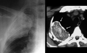
The inside of the mass has a weird appearance with whorls of calcification and areas of decreased opacity. The CT appearance is practically pathognomonic of oleothorax (plombage).
Instillation of oil in the extrapleural space (oleothorax) was used to collapse the lungs facilitating healing of TB cavities. This method was abandoned in the early fifties after the discovery of effective antimicrobial therapy.
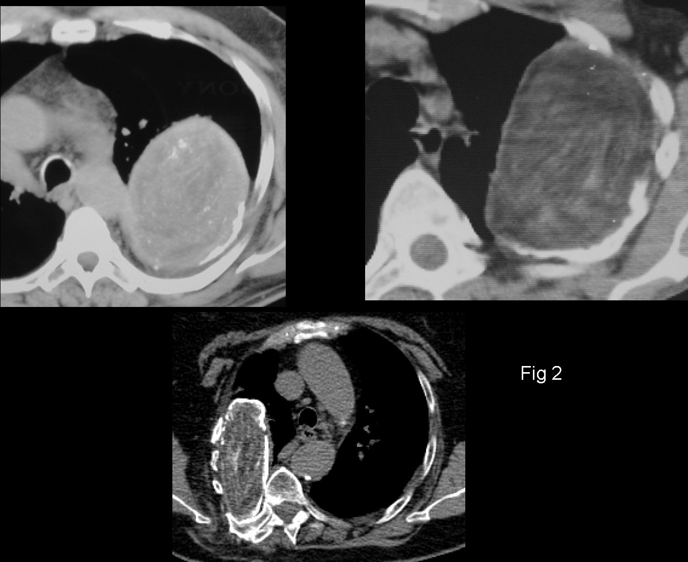
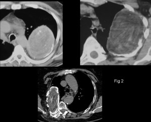
Patients with oleothorax are still seen and it is important to recognize the imaging findings to avoid misdiagnosis. All of them have the same appearance: extrapulmonary mass in the upper chest with absence of the 4th-5th rib (removed to instill the oil). The calcification and the whorly appearance of the mass are nearly pathognomonic. Fig. 2 shows three more cases, one of them seen two years ago.
Teaching point: when confronted with a weird image, always remember to ask the patient for a previous surgical procedure. If he/she does not remember, look for a surgical scar.