Dear Friends,
Muppet gratefully acknowledges the contribution of his good friend Dr. Manel Escobar who discovered and diagnosed the following case:
An 11-year-old girl with migrating bone pain for the last year, with normal radiographs, now presents with one month of low-degree fever. Chest and abdominal CT were unremarkable, except for the pelvic findings.
Diagnosis:
1. Fibrous dysplasia
2. Langerhans cell histiocytosis
3. Lymphoma
4. None of the above
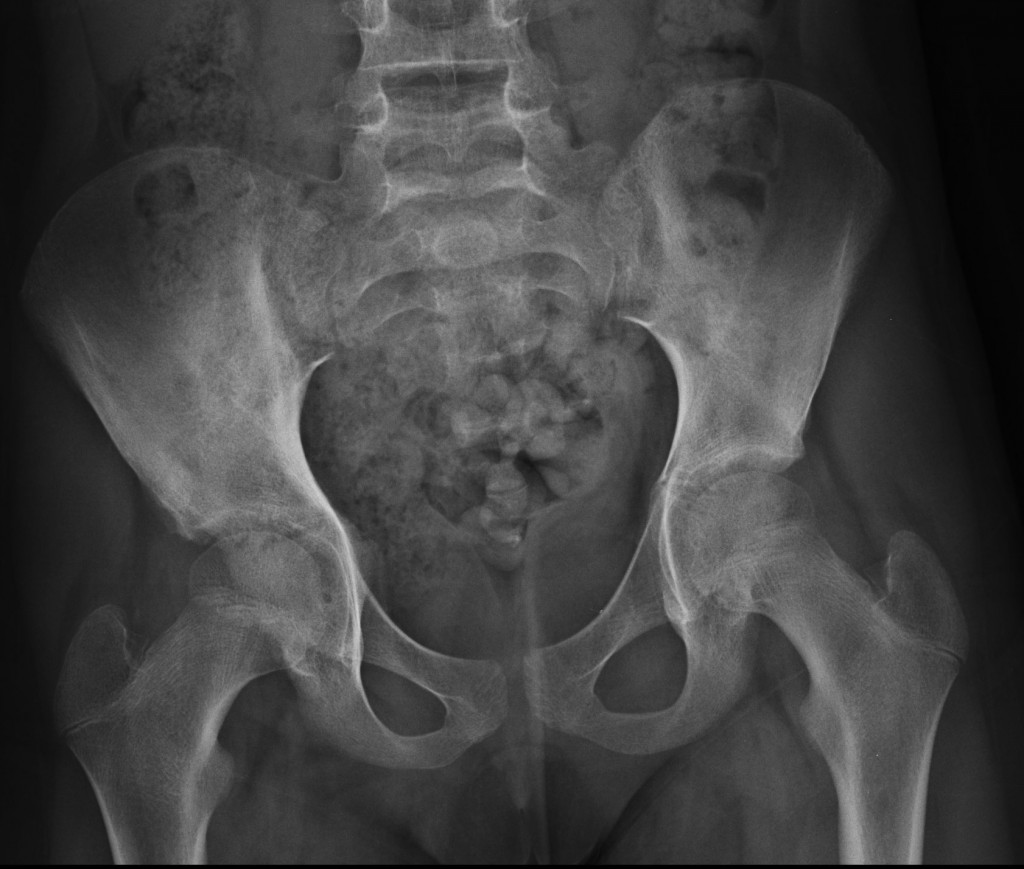
11-year-old girl, AP pelvis
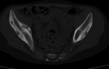
11-year-old girl, axial CT
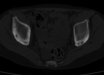
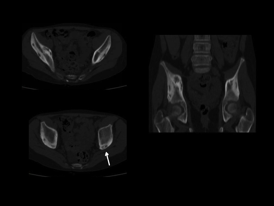
11-year-old girl, axial CT1
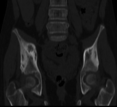
11-year-old girl, corinal CT
CT shows lytic lesions in both iliac bones with a thick periosteal reaction. A sequestrum is seen in the left side (arrow). The presence of a sequestrum and thick periosteal reaction suggests a chronic osteomyelitis. The clinical and imaging findings in this child fit an uncommon disease called chronic recurrent multifocal osteomyelitis (CRMO), which was the final diagnosis in this case.
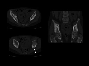
CRMO is an inflammatory process, which affects children. It is a nonbacterial osteomyelitis and typical imaging findings include lytic and sclerotic lesions of bones. Radiologists are usually important in determining the right diagnosis. In this particular case, findings were discovered by the radiologist and the correct diagnosis suggested.
Teaching point: Muppet is not an expert in bone imaging and does not dare to give advice. But he learned a lesson: from now on, chest imaging only!