Dear Friends,
Muppet promises never again to show a bone case. He has been severely reprimanded!
Back to good ol’ chest imaging:
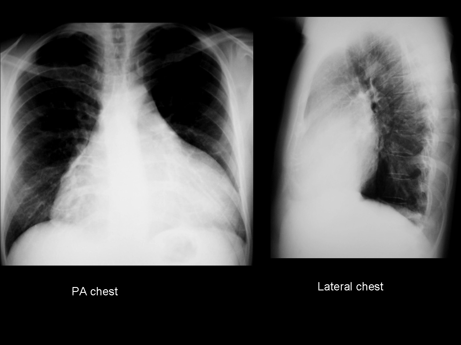
29-year-old male heavy smoker. Admitted with mild dyspnea.
Diagnosis:
1. Mediastinal tumour
2. Dilated cardiomyopathy
3. Pericardial effusion
4. None of the above
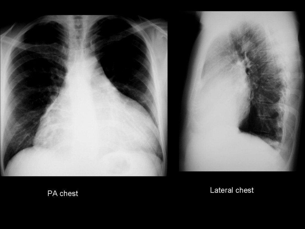
29-year-old male, heavy smoker
Click here for the answer to case #21
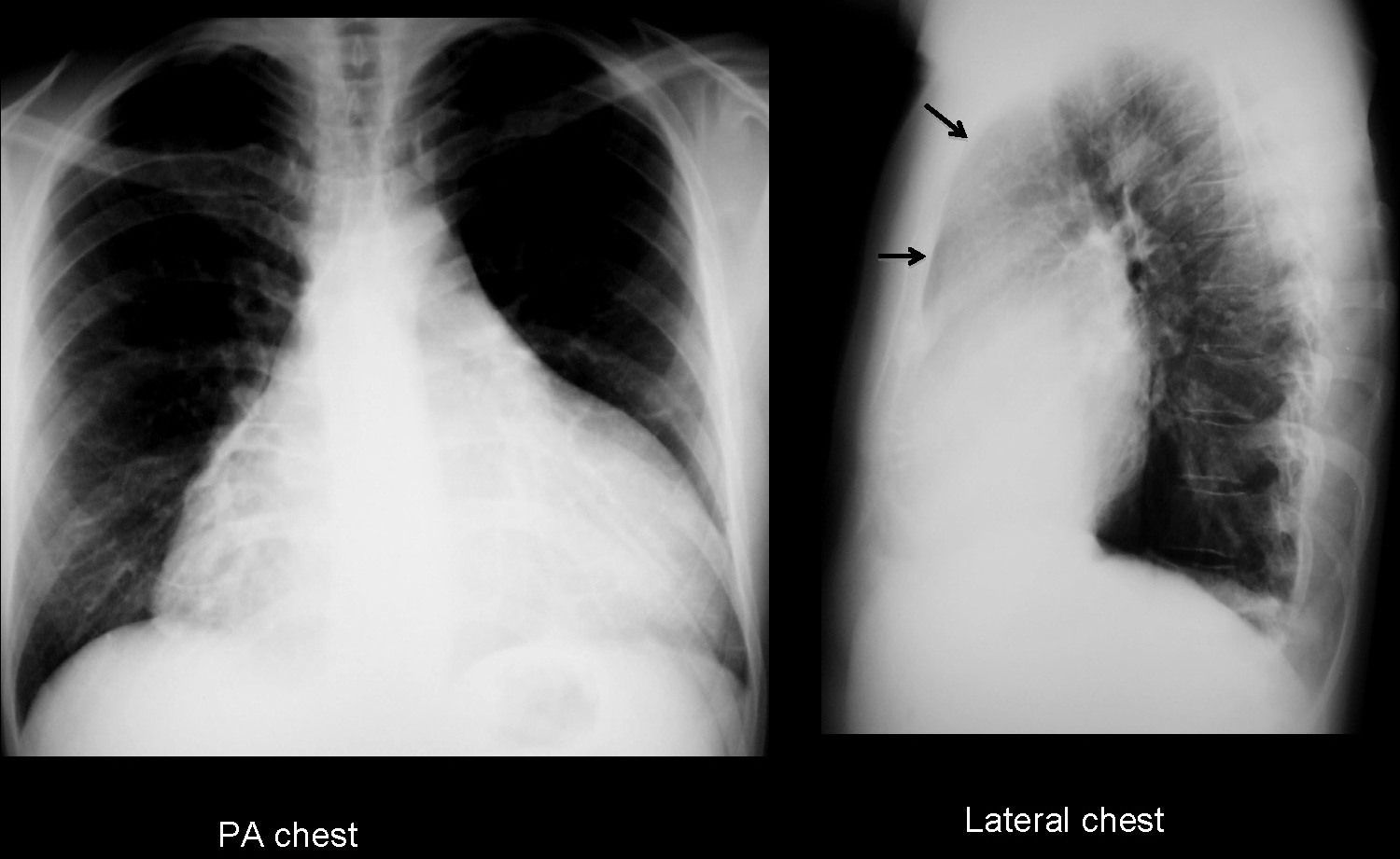
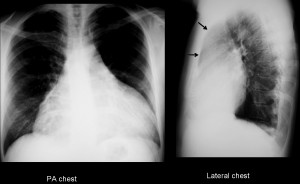
PA chest film shows apparent global enlargement of the cardiac silhouette, with no engorgement of the lung vessels. At first sight, the appearance indicates pericardial effusion. However, the clue to the correct diagnosis lies in the lateral film, which shows occupation of the anterior clear space (arrow), which should be blacker in a 29-year-old person. This finding suggests an anterior mediastinal mass.
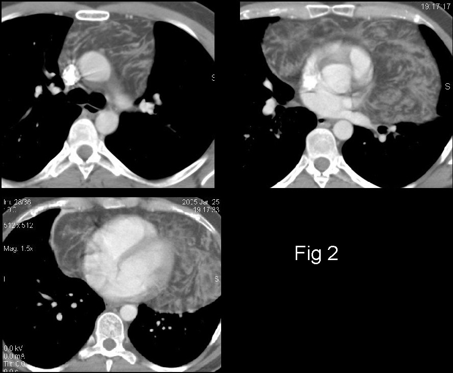
CT confirms the presence of a fatty mass originating in the anterior/superior mediastinum that extends caudally and wraps itself around the heart (fig 2). Thymic masses occasionally grow caudally, appose themselves around the heart and simulate cardiomegaly in the chest radiograph.
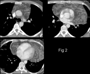
Final diagnosis: thymolipoma, surgically proved.
Teaching point: always look at the anterior clear space in the lateral chest radiograph. You may find pathology not visible in the PA radiograph.