Dear Friends,
Muppet strives to give satisfaction (like Jeeves) and presents you with an easy case:
Fifty-one-year-old male smoker with moderate cough, no fever.
Diagnosis:
1. Metastases
2. Carcinoma of the lung
3. Allergic aspergillosis
4. None of the above
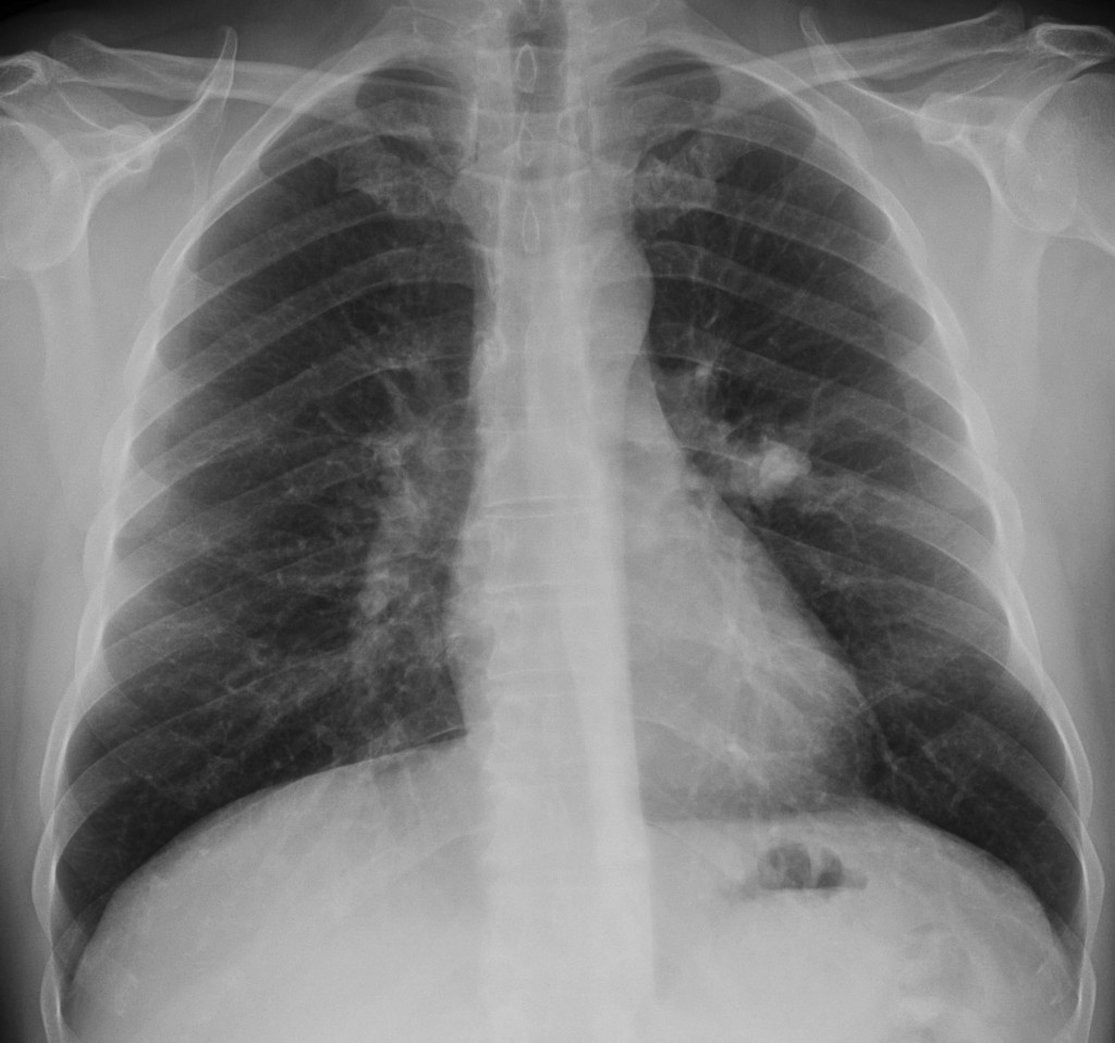
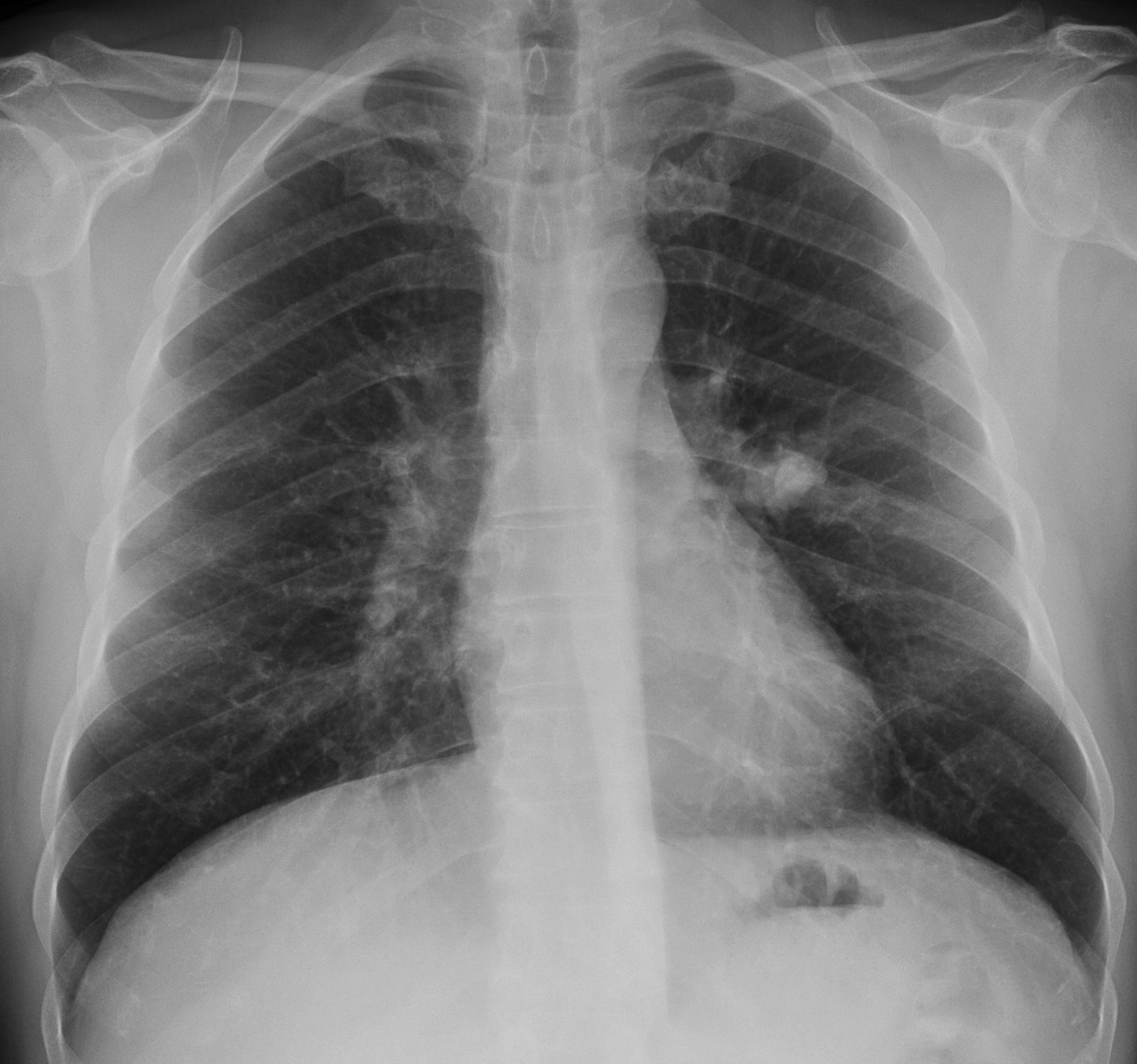
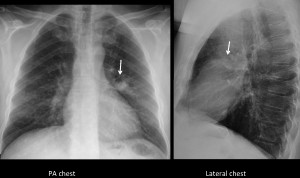
51-year-old male smoker, PA chest
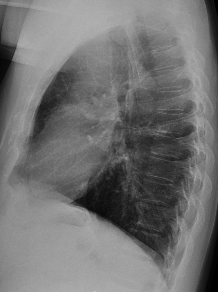
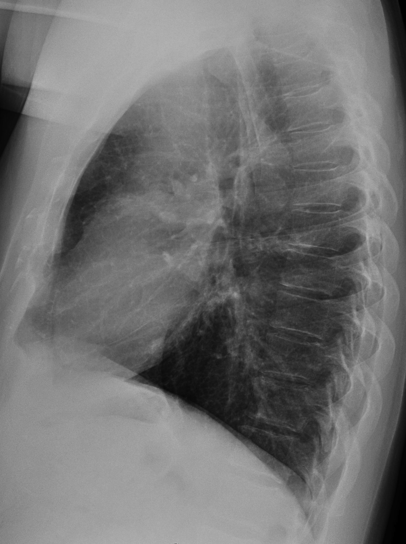
51-year-old male smoker, lateral chest
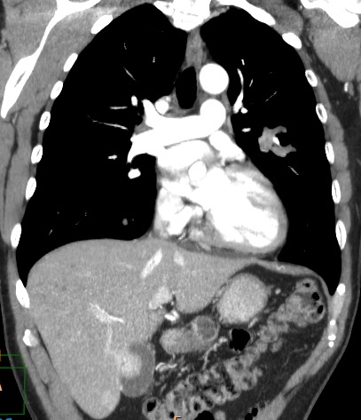
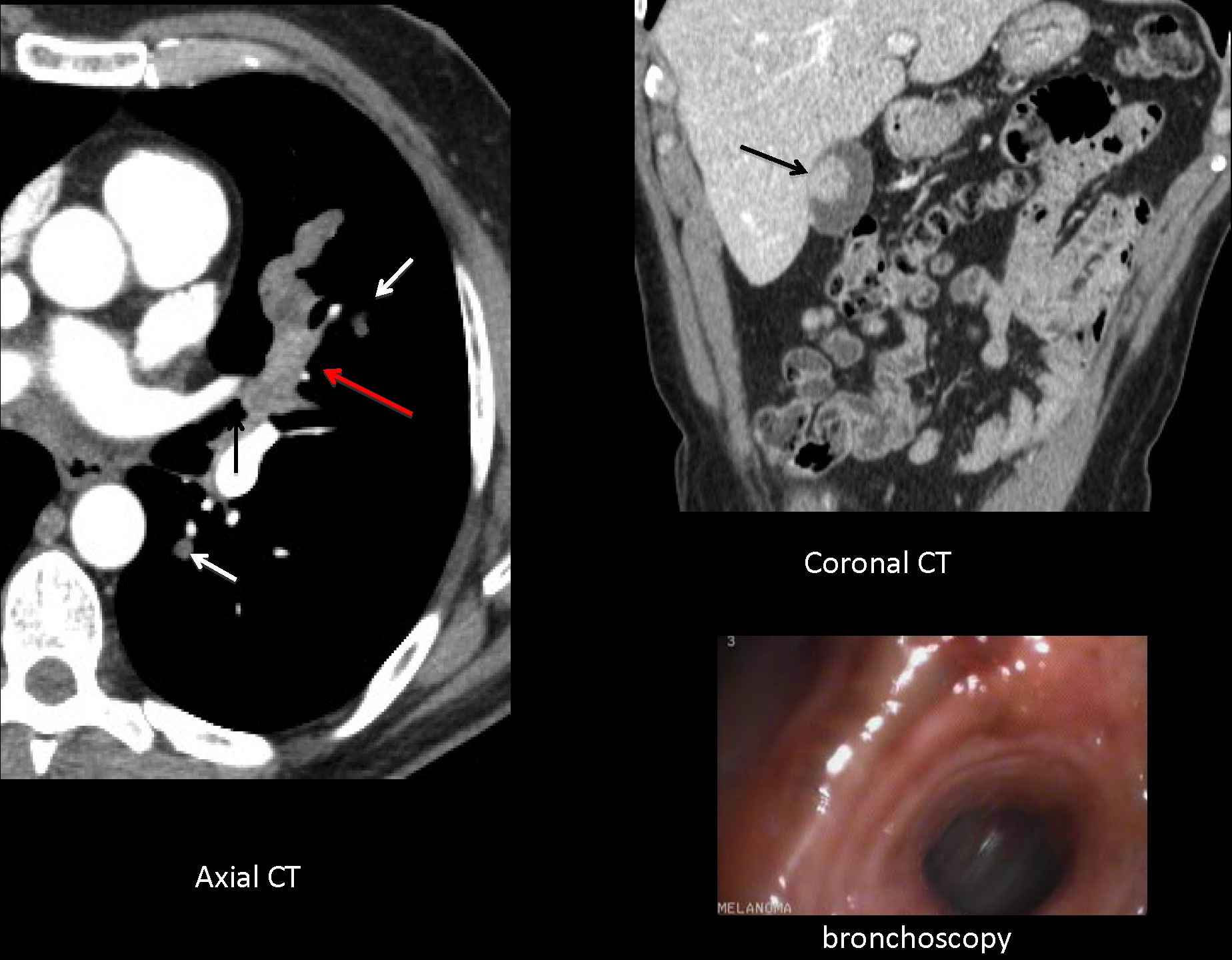
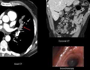
51-year-old male smoker, coronal CT
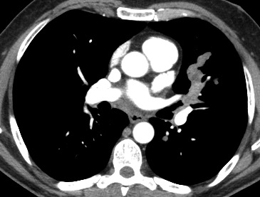
51-year-old male smoker, axial CT
Click here for the answer to case #22
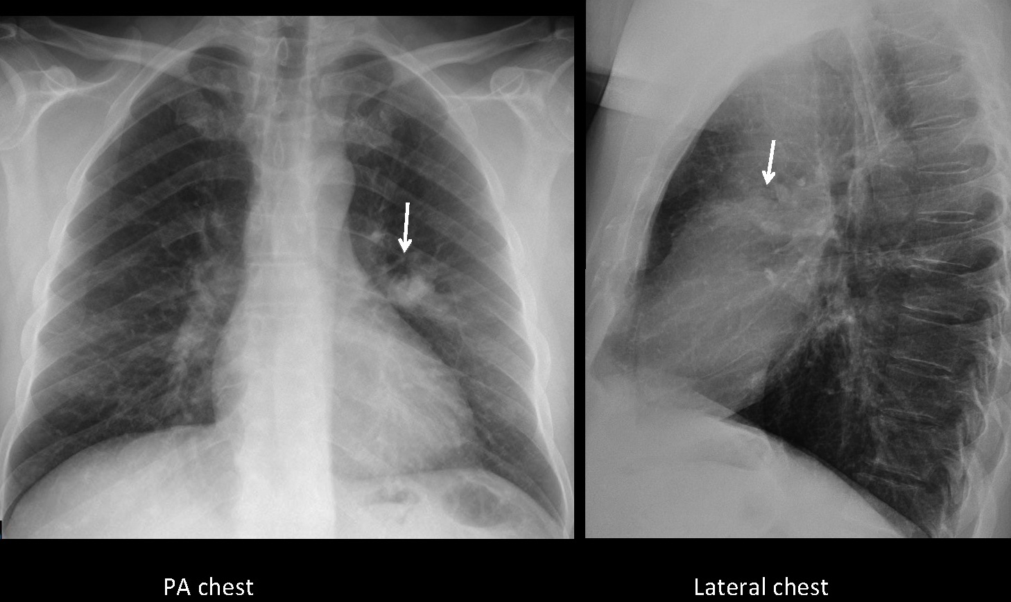
PA and lateral chest show an opacity in the left mid-lung field (arrows). Its shape in the lateral view suggests mucous impaction, confirmed with enhanced axial CT (red arrows). Axial CT also shows an endobronchial lesion (arrow), as well as two small lung nodules (arrows). Coronal CT shows an enhancing nodule inside the gallbladder (arrow).
The combination of an endobronchial lesion, pulmonary nodules and a gallbladder mass suggests widespread metastases, with a high probability of melanoma as the primary lesion (it has been described and Muppet has seen several cases). Bronchoscopy confirms the melanotic lesion in the bronchus. Final diagnosis: widespread metastases from melanoma.
Congratulations to all who made the diagnosis, especially to Albert, who contributed with an excellent discussion.
Teaching point: don’t forget the basic principles (satisfaction of search/sex). Once the abnormality is identified, keep looking. This is an easy case if you gather together all the information and don’t choose the easy way out