Dear Friends,
Muppet is very happy with your progress and wants to test you with an unusual case.
A 57-year-old male with liver carcinoma. No chest symptoms.
What are the densities in the upper lung and mediastinum?
1. Veins
2. Arteries
3. Lymph nodes
4. Esophagus
Warning: this is a difficult case!
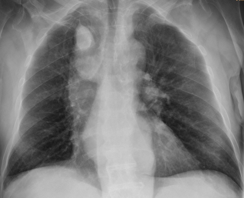
57-year-old male, AP supine chest
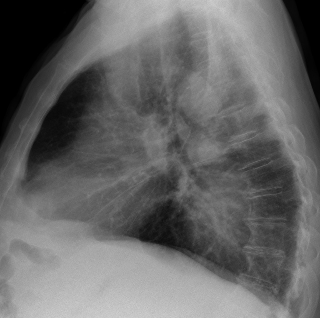
57-year-old male, erect lateral chest
PA chest shows a tortuous line in the left side of the mediastinum (arrows), as well as an azygos fissure (arrows) with an enlarged azygos vein (white arrow). The lateral view shows a prominent azygos (arrow) and an ill-defined haziness in the lower middle mediastinum (white arrows).
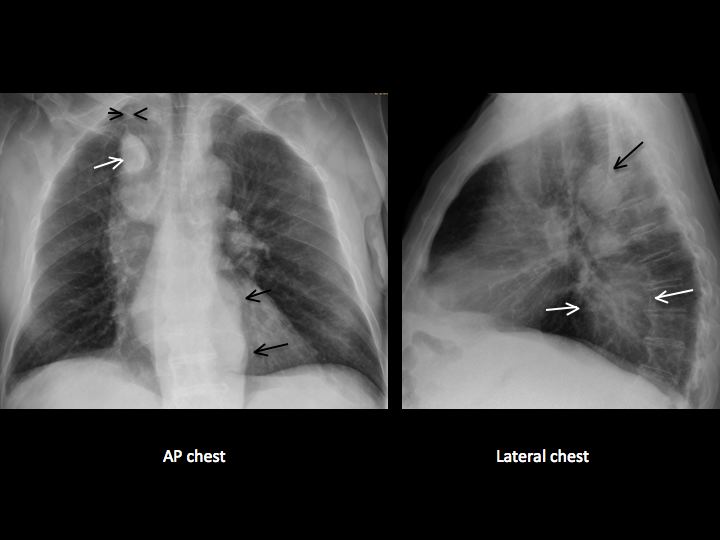
The findings suggest an enlarged azygos/hemiazygos system accompanied by an azygos lobe. Findings are confirmed by coronal and sagittal CT, which show the enlarged azygos and hemiazygos veins (arrows). Axial CT shows the azygos lobe (arrow).
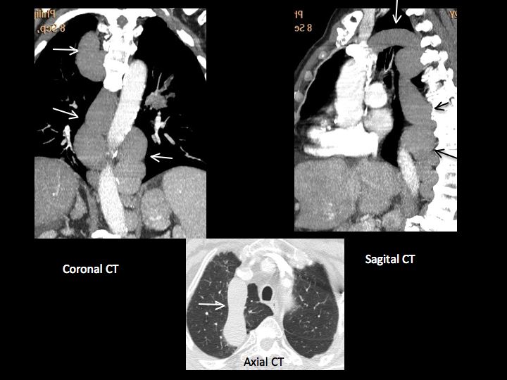
Muppet was surprised because these findings were not related to liver disease or varices. The patient had a congenital absence of the inferior vena cava with azygos continuation, combined with an azygos lobe. Muppet’s alter ego published this rare combination a long time ago.
Teaching point: if we pay careful attention to the findings and make a good semiologic reading, it is possible to diagnose unusual conditions, as proven by the many correct answers to this case. Congratulations!


Lymph nodes.
I think we can see occupation the inferior medium mediastinum (convexity of the pleuroacigoesophaeal recess and double left cardiac contour not corresponding to the aorta neither the paravertebral line); finding that suggest ESOPHAGEAL VARICES.
Then we can see an ACIGOS VEIN markedly dilated also because of the portal hypertension and the normal anastomsis between esophageal veins and acigos-hemiacigos system.
The funny thing of the case is to explain the “intrapulmonary” oppacity in the upper right lobule. I will try: that also correspond to the ACIGOS VEIN because there is an ACIGOS LOBE as an anatomical variant.
Summarizing:
Extensive esophageal varices and acigos dilatation with a “tricky acigos lobe”.
I think it isn’t possible to establush the cause of the acygos dilatation. If you see it isolated in asimptomatic pacient I would think of IVC agenesi; in this pacient I considered IVC trombosis (posibly tumoral) and portal hypertension “per se”. Taking in account the fact that I believe there are extensive esophageal varices I chose portal hypertension as common cause for both, but cant ensure IVC trombosis exist.
Enlarged paraspinal spaces (displacement of the lines bilaterally) + azygos cross in accesory fissure. Middle mediatinum is enlarged to the right probably because of the SVC.
D+: Inferior vena cava thrombosis (probably complete) with collaterisation through hemiazygos-azygos system.
Now that’s extreme radiology (wow).
Absence of IVC with collateral circulation and dilatation of acygos vein (inside an acygos lobe)
And the answer is Nº1 Veins
the well-defined opacity with smooth margins on the upper right lobe (vascular structure) seems to be inside a cavitary lesion (rasmussen aneurysm? puzzling, without any chest symptoms). On the later view there is a large area of increased attenuation with ill defined margins between heart and dorsal spine.
Thank you for this case but quite difficult Dr Caceres!
Partiamo dal dato clinico:il tumore epatico.Una delle possibili complicanze è l’invasione tumorale di qualche vena epatica: l’occlusione lenta e cronica porta ad ipertensione portale , con sviluppo di circoli di compenso ; un circolo collaterale è rappresentato dalla V Azigos, che pertanto si dilata. siamo di fronte allora ad una s: di Budd.chiari, asintomatica, perchè lentamente occlusiva , compensata dai circoli collaterali, senza epatomegalia-ittero ed ascite, e solo circoli di compemso.
Mr. Muppet, the hardest case ever, congratulations.
Hard cases are fun. Make us think (a forgotten passtime)
Azygos vein dilatation in the accesory azygos vein lobe (due to the portal hypertension? secondary to the portal vein thrombosis?). I am still working on mediastinal findings. Warm greeting to Mr. Muppet 😉
Arteries
hmmm… i can’t stop thinking about this case…
1. in the lungs – there are changes of peribronchovascular interstitium, venous stasis, fibrotic deformations and maybe infiltration of S10 ir the right.
2.In the RUL – accesory fissure and v.azygos lobe.
3.wide and intensive the mediastinum shadow (middle, inferior) – probably because of the SVC and esophageal pathology.
4.in the right hilum there is intensive shadow, which can be lymph nodes, dilated venous or maybe even an esophageal diverticulum.
mostly veins
prominent azygos vein with accessory azygos fissure probably due to thrombosis of IVC, as the patient is suffering from Liver malignancy. ANS is No. 1
Azygos vein and azygos lobe!
azygos vein and esophageal diverticulum
Il caso era ifficile, ma gli aiuti dell’illustre collega, proponendo le diverse opzioni, lo ha reso più facile.
you love this? baby convertible cribs online shopping pMoUXdHJ http://www.best-babycribsforsale.com/category/modern-and-luxury-baby-cache-cribs/
sell brian cushing red jersey , for special offer TmYlGTTc http://houston-texans-jersey.net/brian-cushing-limited-black-game-jerseywhite-elite-men-jerseyred-youth-women-jersey/