Dear Friends,
Muppet is very happy to have reached the 25th case and hopes for twenty-five more!
Forty-nine-year-old woman with persistent cough for the last two months.
Diagnosis:
1. Carcinoma
2. Tuberculosis
3. Bronchiectasis
4. None of the above
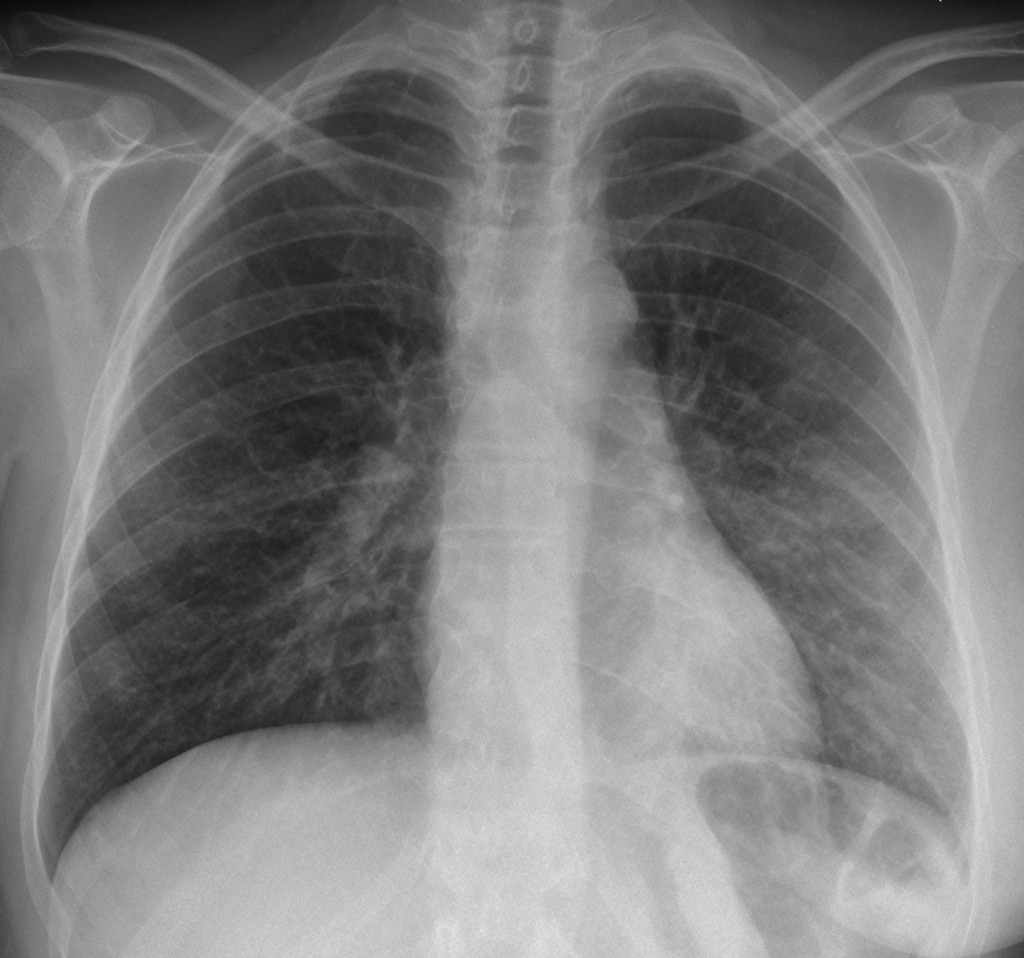
49-year-old woman, PA chest
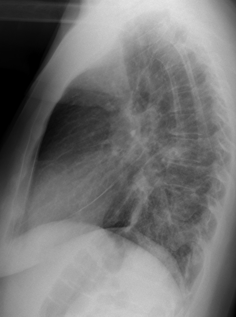
49-year-old woman, lateral chest
Click here for the answer to case #25
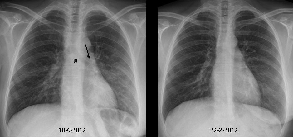
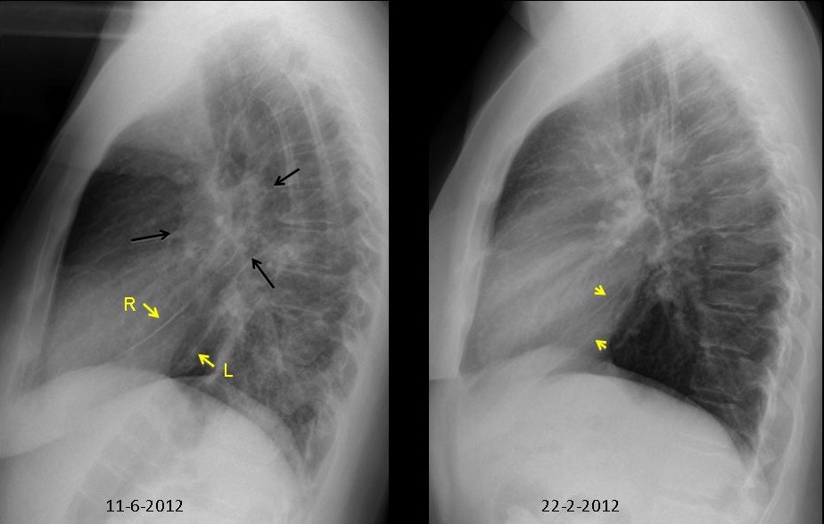
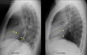
PA chest shows non-descript opacity in LLL with moderate downward shift of the left hilum (arrow), partially hidden behind the heart shadow, accompanied by a vertically oriented left main bronchi (arrow). The lateral film shows a hilar donut sign (arrows) and posterior displacement of the left major fissure (yellow arrows). These signs indicate moderate volume loss of LLL and they are evident when compared with radiographs taken four months earlier.
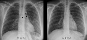
In a patient with mastectomy (absent right breast in PA film), the most likely diagnosis is either metastatic disease or primary bronchial tumour.
CT shows peribronchial metastatic infiltration of LLL bronchus with lymphangitis and mediastinal and hilar lymph nodes with hypodense centre, probably due to chemotherapy. Previous CT was normal.
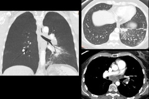
Final diagnosis: metastatic carcinoma of the breast.
Teaching point: this case illustrates the main findings of LLL collapse. Conventional radiography offers a lot of information if we look for it (see Marius’ interpretation above).