Dear Friends,
This case has been provided by Dr. Oscar Persiva, a former resident and good friend (even though he is a fan of Real Madrid).
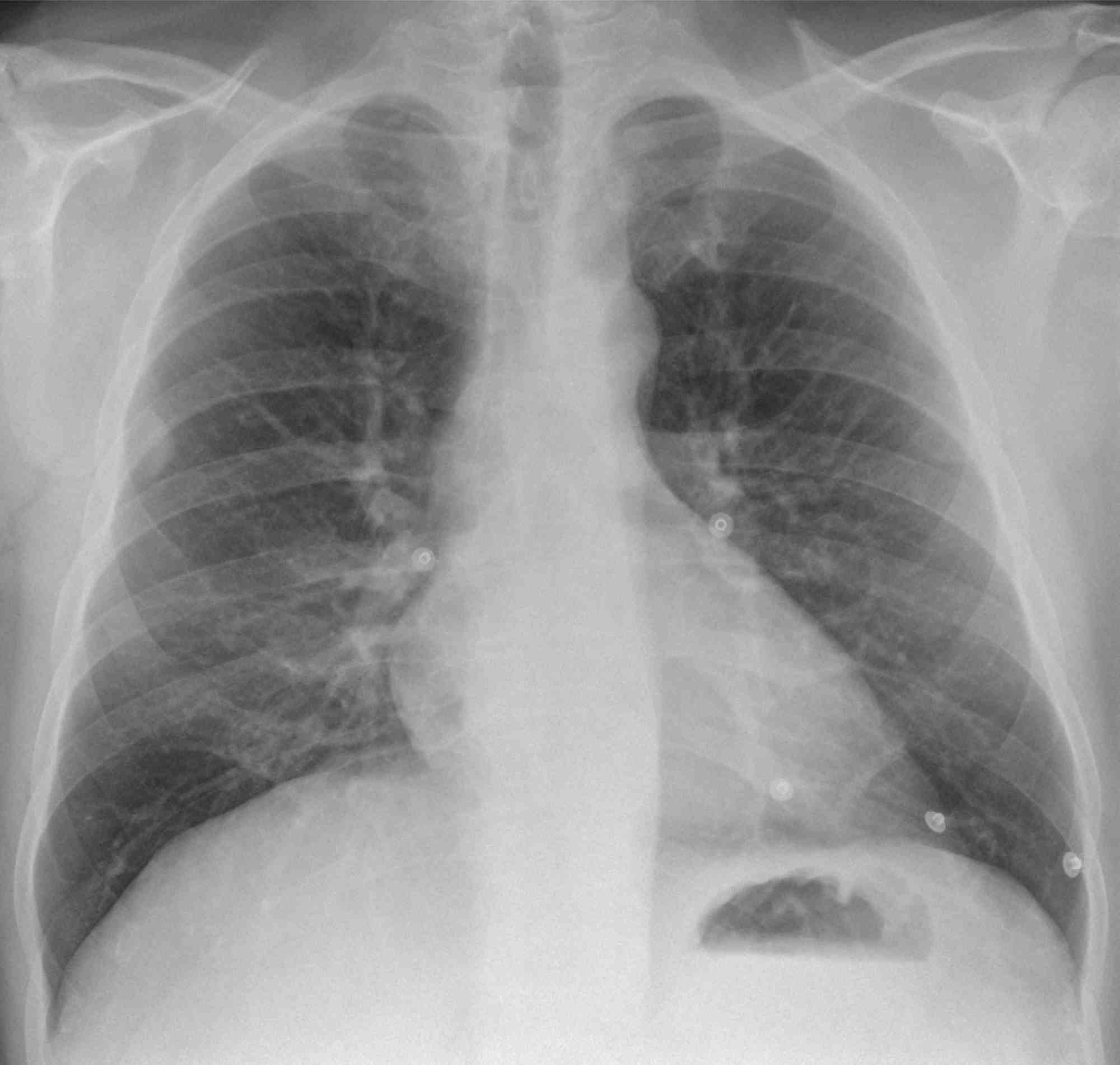
The case is a 38-year-old man who came to the Emergency Room with moderate chest pain. Chest radiograph and CT shown.
Diagnosis:
1. Pleural metastases
2. Neurogenic tumour
3. Fibrous tumour of pleura
4. All of the above
38-year-old man, PA chest
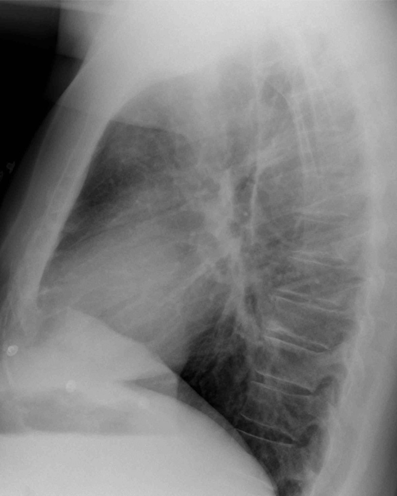
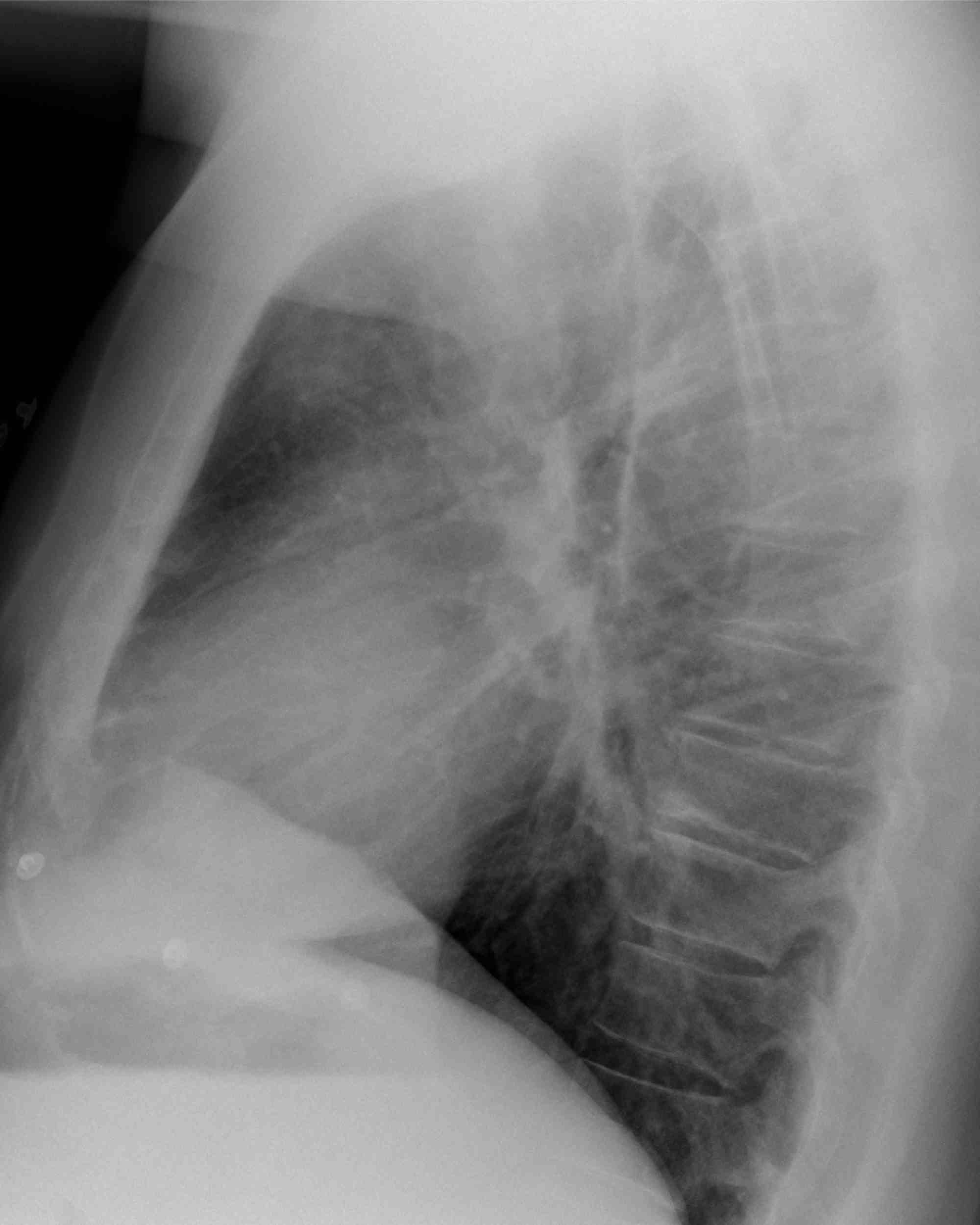
38-year-old man, lateral chest
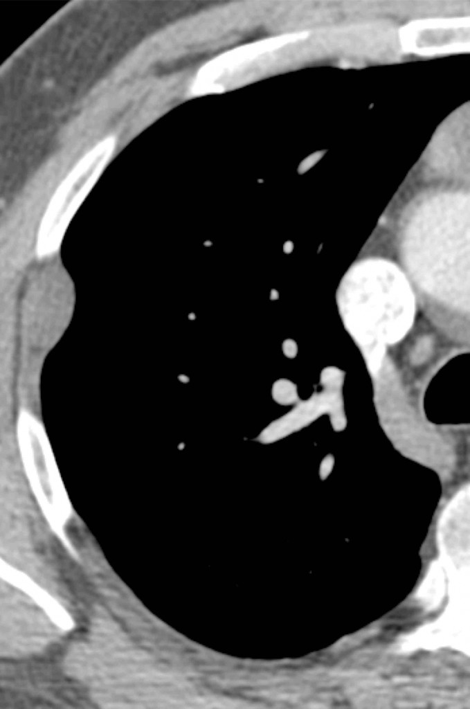
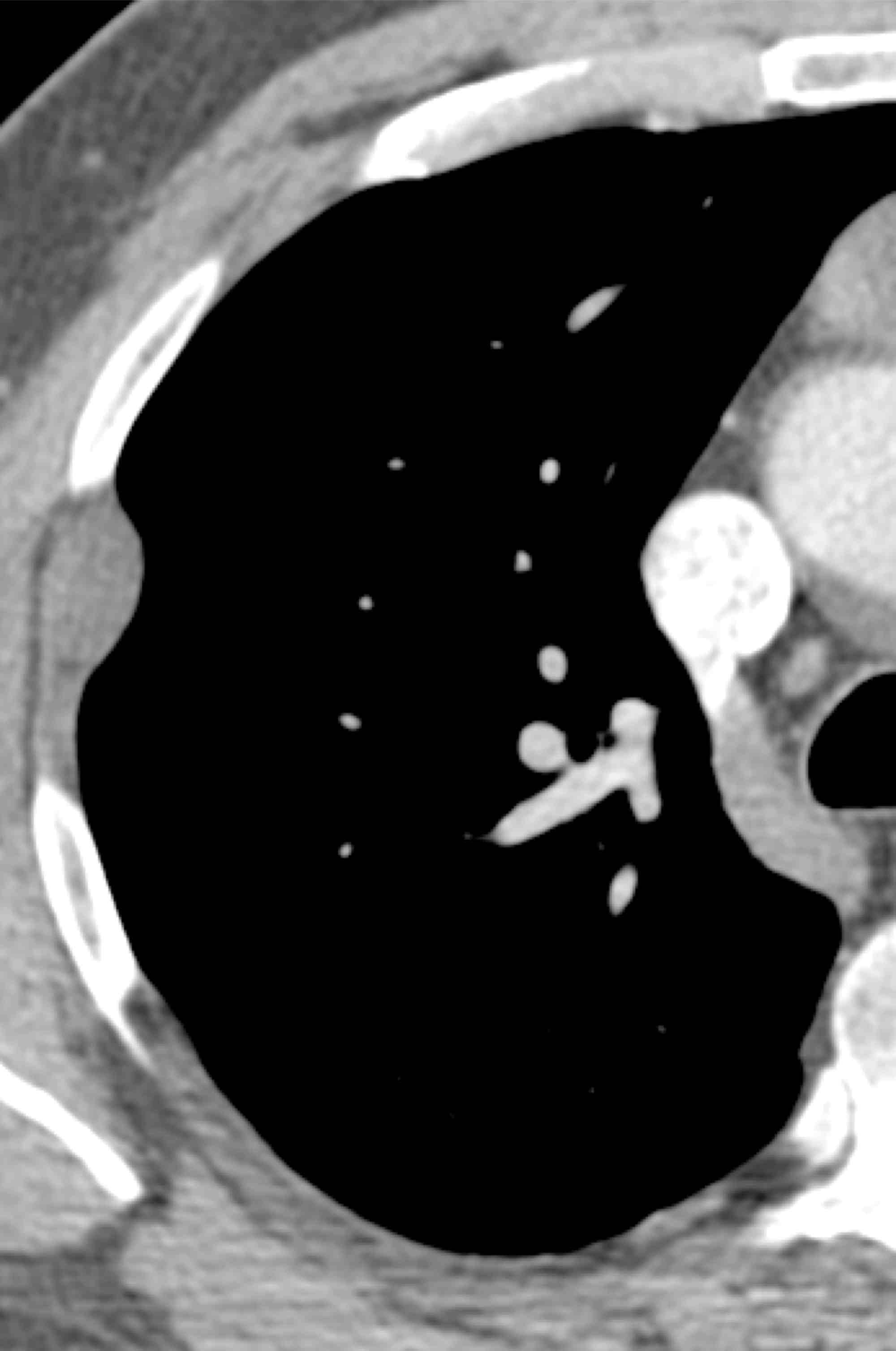
38-year-old man, axial CT
Click here for the answer to case #27
PA chest film shows a peripheral nodule (arrow). The lateral film shows increased opacity in the anterior clear space (arrow). Axial CT confirms an extrapulmonary nodule (arrow), as well as a rounded anterior mediastinal mass (red arrow).
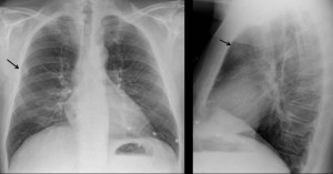
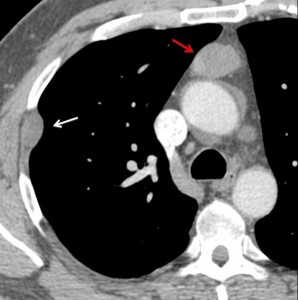
This combination suggested to the Muppet that this was a malignant thymoma with pleural metastases, as thymomas metastasize along the pleural layers. However, a thymoma with a coincidental chest wall mass could not be excluded.
Therefore, the correct answer should have been: ‘4. All of the above’
The patient was operated on and the final diagnosis was benign thymoma with chest wall schwannoma. The Muppet, thinking he was infallible, is now quite ashamed of himself.
Teaching point: Don’t forget the satisfaction of search/sex! In this patient the discovery of a mediastinal mass in the lateral chest raised interesting diagnostic questions.
Point 2: Always look at the anterior clear space on the lateral film.