Dear friends,
As summer is here and the weather is hot, Muppet wants to show you an easy case:
Preoperative chest for urethral stenosis in a 26-year-old male.
Diagnosis:
1. Pleural mesothelioma
2. Lung tumor
3. Loculated fluid
4. None of the above

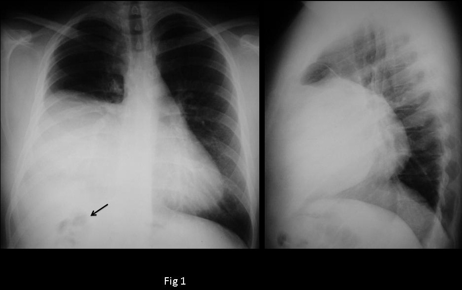
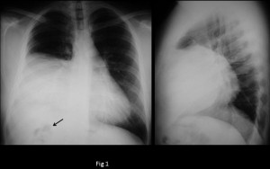
26-year-old man, PA chest

26-year-old man, Lateral chest
Click here for the answer to case #28
PA and lateral chest show an extrapulmonary opacity in the right lower lung field. There is a loop of bowel in the right upper quadrant (arrow), which may be due to Chialiditi’s syndrome or elevation of the colon, secondary to diaphragmatic hernia.
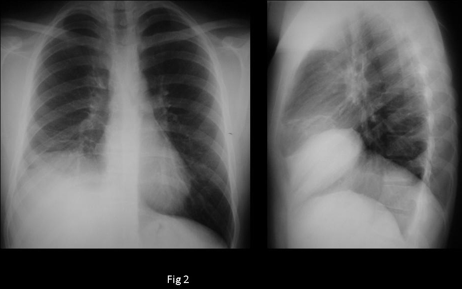
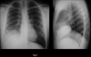
Muppet considered that hernia was a good possibility and investigated the clinical history: urethral stenosis was secondary to rupture of urethra and bladder neck as result of a motorcycle accident four years earlier. Chest films two years earlier (Fig 2) showed apparent elevation of the right hemidiaphragm, which was not reported.
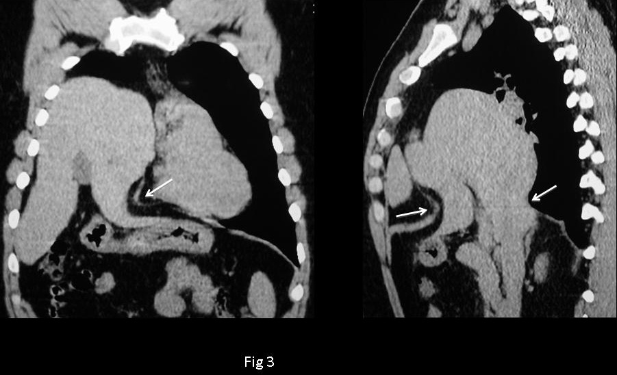
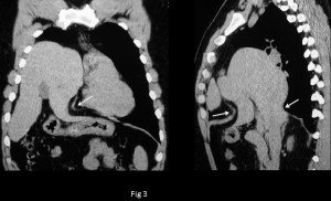
These findings increased the suspicion of traumatic diaphragmatic hernia and led to a CT, which showed a rent in the diaphragm (Fig 3, arrows) with herniation of the liver. Hernia was surgically repaired; Muppet saved a life and was very proud of himself!
Teaching point: any lesion in close contact with the diaphragm may arise from below. In case of doubt, it is imperative to perform a CT.