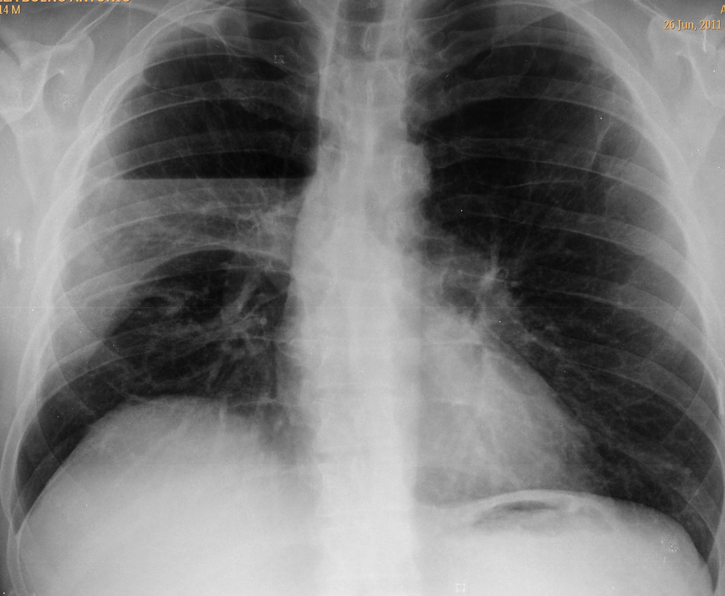
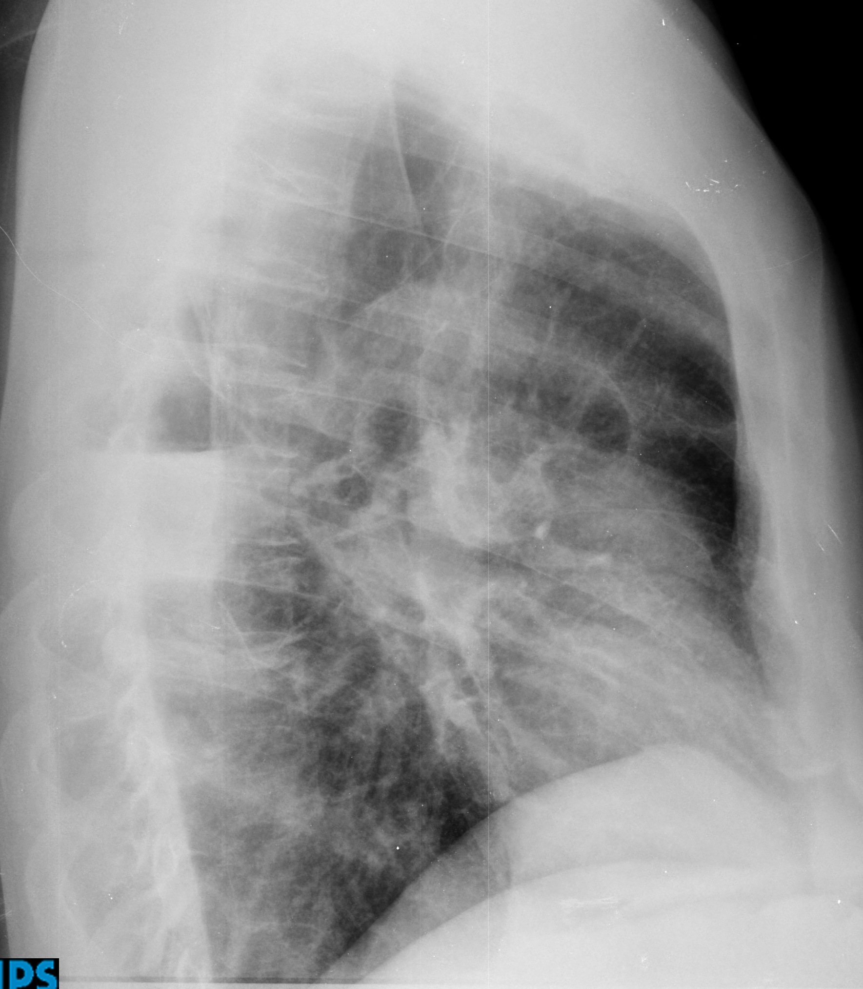
During August, Muppet would like to discuss some basic principles of interpretation. The first case is a 46-year-old male with neurofibromatosis and bullous emphysema, who presents with fever.
1. Hidroneumothorax
2. Infected bulla
3. Lung abscess
4. None of the above
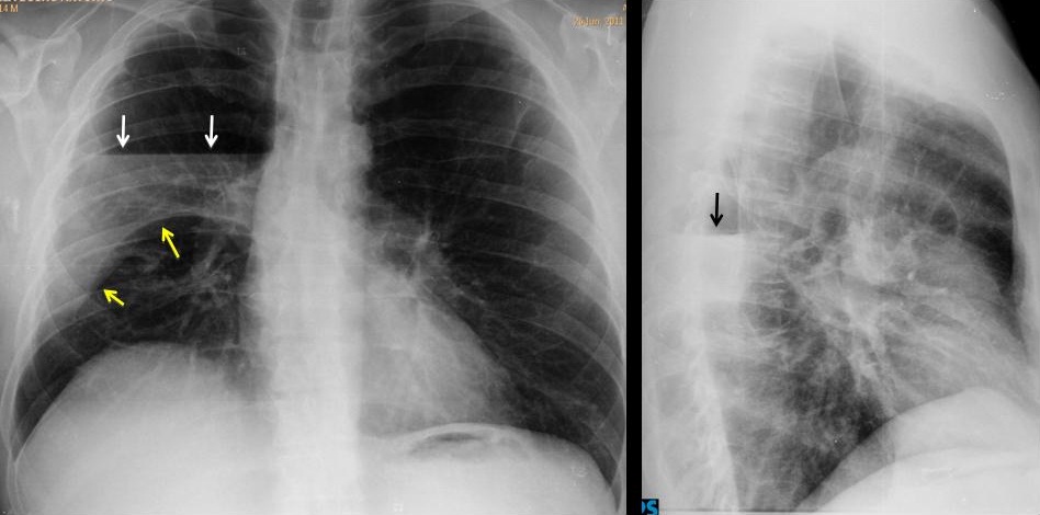
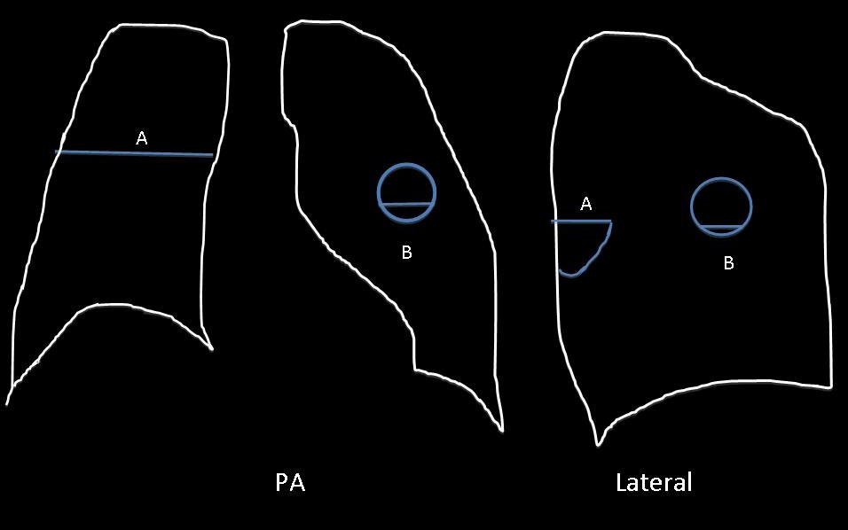
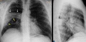
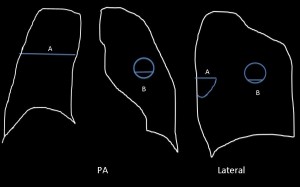
PA film shows an air-fluid level, which crosses the right hemithorax (arrows). The same air-fluid level, considerably shorter, is seen in the lateral view, (arrows). This appearance is very suggestive of a hydropneumothorax (A), as opposed to a pulmonary cavity (B), where the air-fluid level is of the same size in both projections (see drawings).The undulated line in the PA chest corresponds to the major fissure outlined by pleural fluid in its superior aspect (yellow arrows).
CT confirms the diagnosis by showing air and fluid in the pleural space (fig 2, arrows). There are numerous bullae, none of which has fluid.
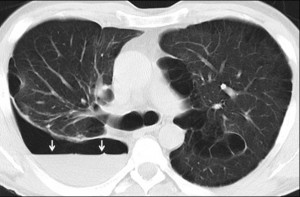
Final diagnosis: hydropneumothorax (empyema, since the patient was febrile)
Teaching point: this case shows the typical features of hydropneumothorax in the plain film:
1. Air fluid levels are of different size in PA and lateral views
2. Lack of upper wall (visible in pulmonary cavities)