Welcome to case number three!
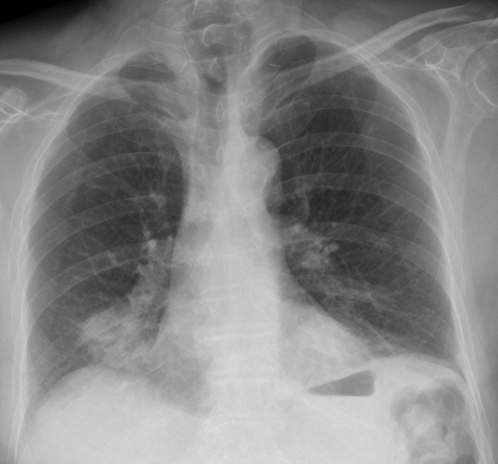
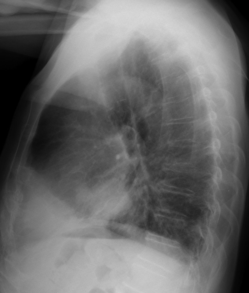
My wife, who works in a primary care centre, sent this case to me. The patient is a 76 year-old male, who was operated on three years ago for carcinoma of the larynx. The PA and lateral radiographs show two nodular lesions in the lower lung fields.
The obvious response is metastatic disease. But the Muppet, trying to impress both of us, suggested an alternative diagnosis.
Can you guess the Muppet’s diagnosis?

76 year-old male with two nodular lesions in lower lung fields (PA)

76 year-old male with two nodular lesions in lower lung fields (lateral)
Click here for the answer to case #3
Answer to case #3
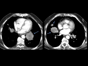
To the eternal shame of the muppet (and myself), my wife was right. The correct diagnosis of case #3 is pulmonary metastases. A CT study showed several necrotic masses in the lower lungs (arrows).

I thought that lower lobe infiltrates in a patient with a probable swallowing disorder made aspiration a good possibility. The muppet went even farther and insisted on the diagnosis of paraffinomas, secondary to aspiration of mineral oil.
Why was I wrong? Because I work in a big University Hospital and try to make fancy, impressive diagnosis. Forgot to follow the KISS standard: Keep It Simple, Stupid.
Teaching point: Make things simple. Dr. Felson used to say: ‘Always consider an unusual manifestation of a common disease rather than a common manifestation of an unusual disease’.


syphilitic lesion or lung tuberculosis
Atelektasis of the Middle lobe
Bronchial carcenoma with atelectasis segment
Because the patient is a smoker, the patient has a bronchial carcinoma in both power lobes. Unfortunate man.
I meant lower lobe, not power lobe.
Sorry, Frank the muppet is more sophisticated! Which does not mean he was right!
second primary lung carcinoma
It’s a fish!
It is not. But it’s a fishy case!
Lung abscess
Bronchial carcenoma with atelectasis segment in right lung, and metastasis to the lymph node left lung
-prominent air shadow to the left of upper trachea -?pharygeal pouch/esophageal diverticulum or post op change. -nodular lesions with lobulated margins in the lower lobes with reticulonodular shadowing. -plate atelectasis adjacent to left hemidiaphragm . Possible diagnoses are :changes of aspiration (mineral oil). Benign lesions like hamartoma/pulmonary manifestations of laryngeal papillomatosis/bronchocele
Good! The muppet likes your diagnosis. What would you do next?
aspiration pneumonia due to diverticulum of esophagus? barium meal and CT of course
aspiration pneumonia due to diverticulum of upper esophagus? barium meal and CT of course
i shall do HRCT chest .and barium swallow.
Excellent answers. You are both right. Congratulations
can it be lymphangitis carsinomatoza ?
it is two pulmonary abscess without entrance to bronch
round atelectasis and encapsulated pleural effusion-anterior C-F sinus is obliterated by effusion and spread it in a major incisura …dif.dg.Renal Ca meta,nodular benign lesions,hamartoma but have no pop corn changes in it,A-V malformation….earlyer radiographics must be checked too 🙂
He had a carcinoma of the larynx so he was probably smoker. The bronchial walls are thickened due to chronic bronchitis. He could also have bronchiectasias that are now filled with mucus. Nevertheless, I´d recommend a non-contrast CT.
Professor thank you once again for challenging case.
Thank you, Anna. I try to do my best