Muppet is feeling mean today and wishes to inflict upon you the following case: a 56-year-old woman with a history of respiratory infections. She was operated on for osteogenic sarcoma of the right leg eight years earlier.
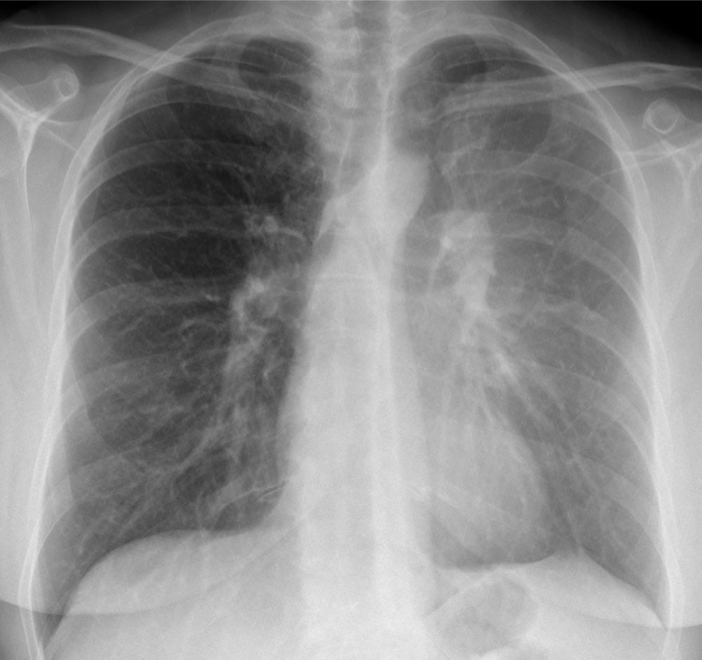
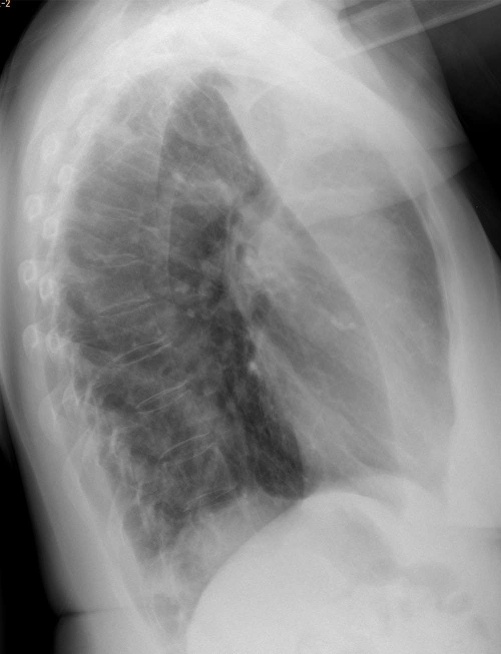
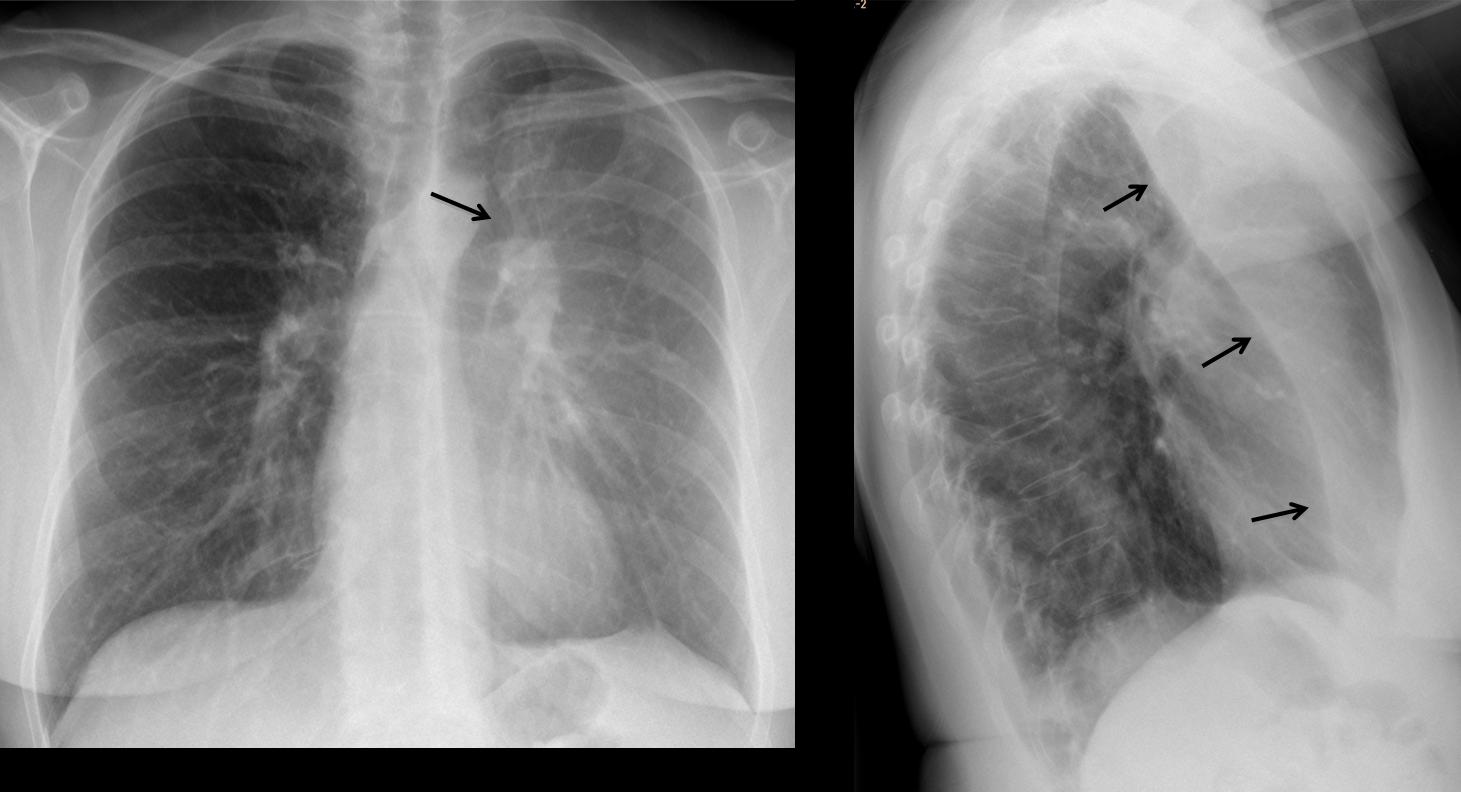
PA and lateral chest show typical signs of collapse of LUL, with haziness of hemithorax, luftsichel (Fig 1, arrow) and marked displacement of the major fissure on the lateral view (Fig 1, arrows).
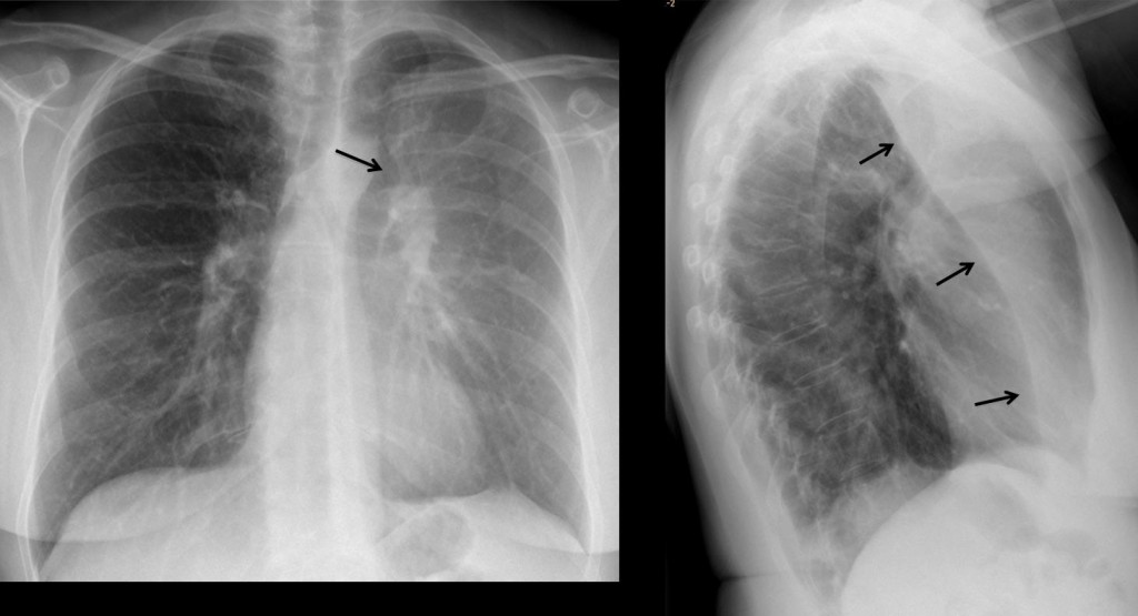
Fig. 1
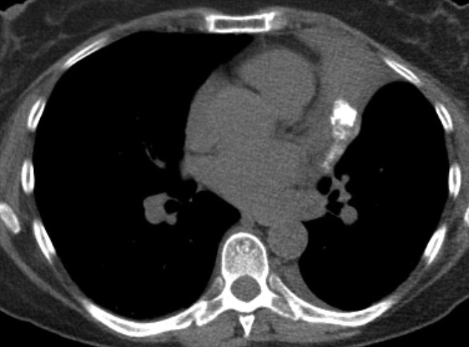
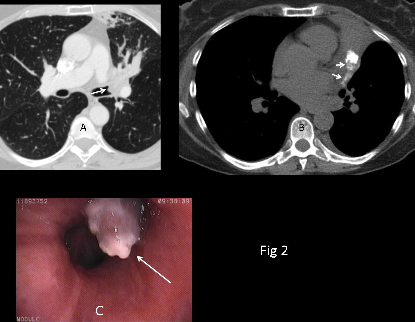
CT demonstrates an endobronchial lesion at the origin of the LUL (Fig 2A, arrow) and heavy calcification along the path of the bronchus (Fig 2B, arrows). Bronchoscopy confirms the endobronchial mass (Fig 2C, arrow).
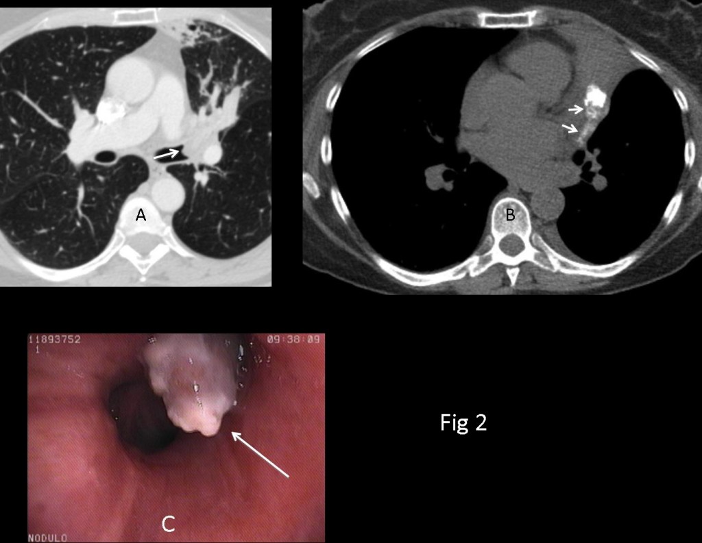
Fig. 2
The most common endobronchial mass is lung carcinoma, but the calcification goes against it. Broncholiths and foreign bodies are not usually located in upper lobes and calcium is better defined. Carcinoid tumour and hamartomas are a possibility. Given the previous history of osteogenic sarcoma, endobronchial metastases should be considered.
Final diagnosis: endobronchial metastases from osteogenic sarcoma.
Teaching point: The most common cause of LUL collapse is first and foremost a carcinoma of the lung. Endobronchial metastases can give a similar appearance and are more common in tumours of the breast, kidney and melanoma although they may occur in any type of tumour, as in the present case.