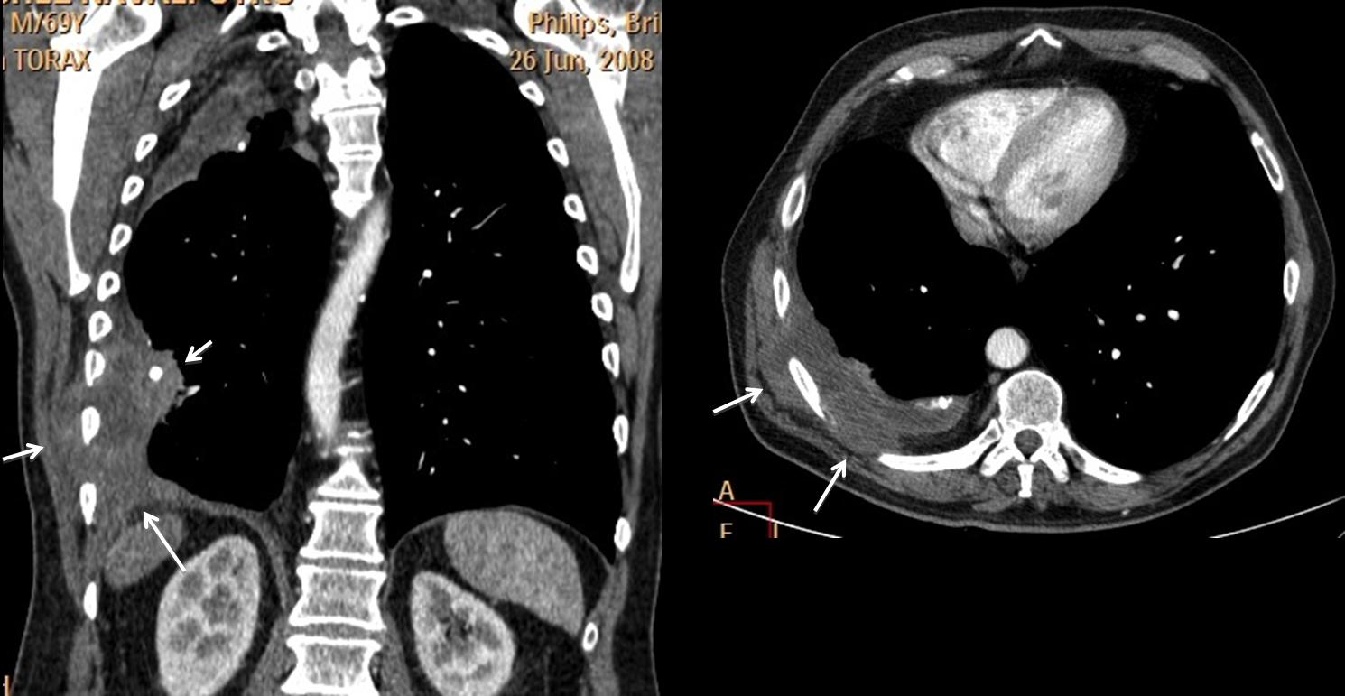
Muppet is feeling good today (love letter from Miss Piggy) and wants to show you an easy case: a 69-year-old male with chest pain and productive cough. TB in his youth.
1. Active TB
2. Empyema necessitatis
3. Pleural abscess
4. None of the above
Dear friends,
I don’t believe that the case is difficult. In fact, if you analyse all the findings, it is reasonably easy.
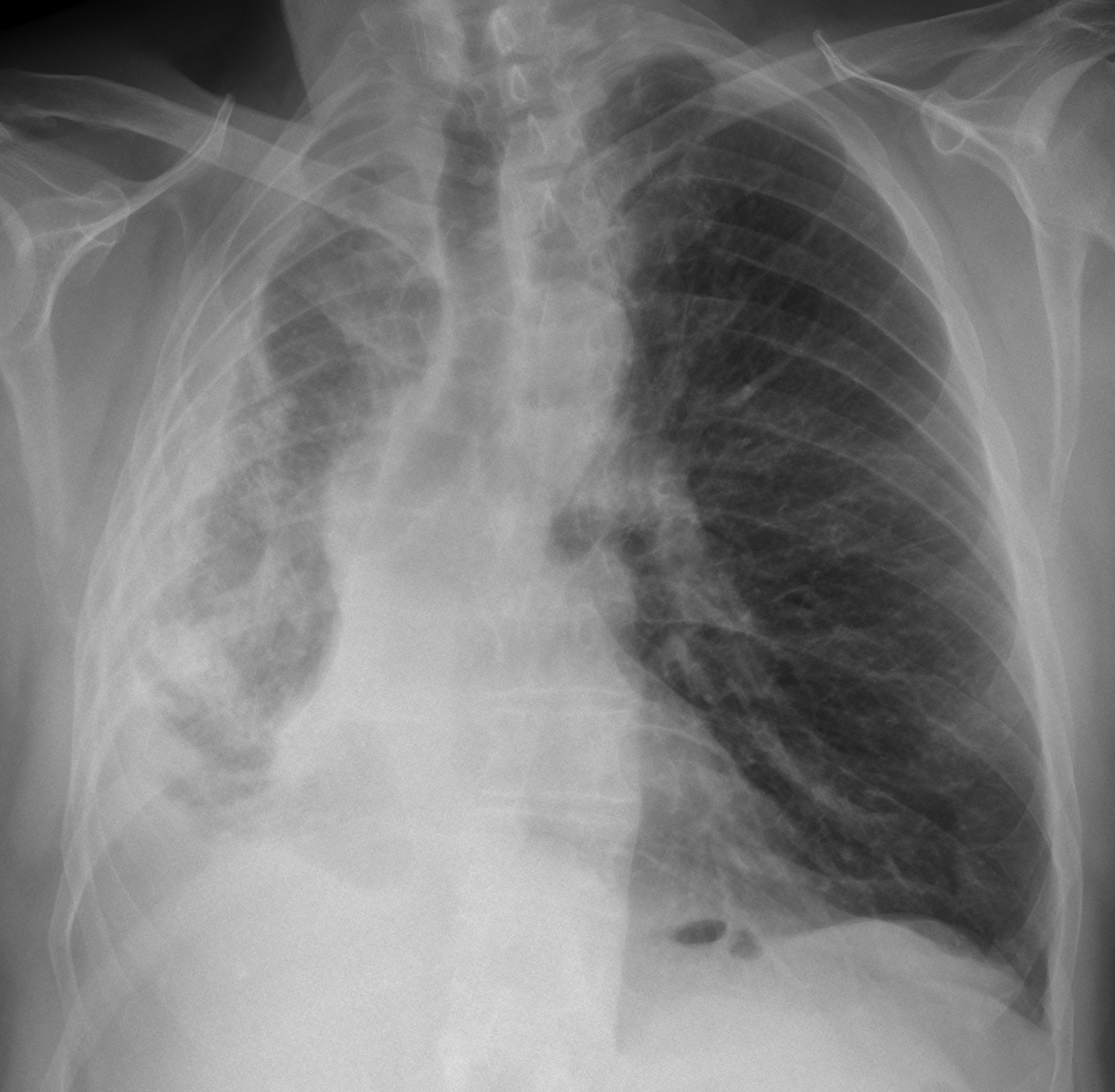
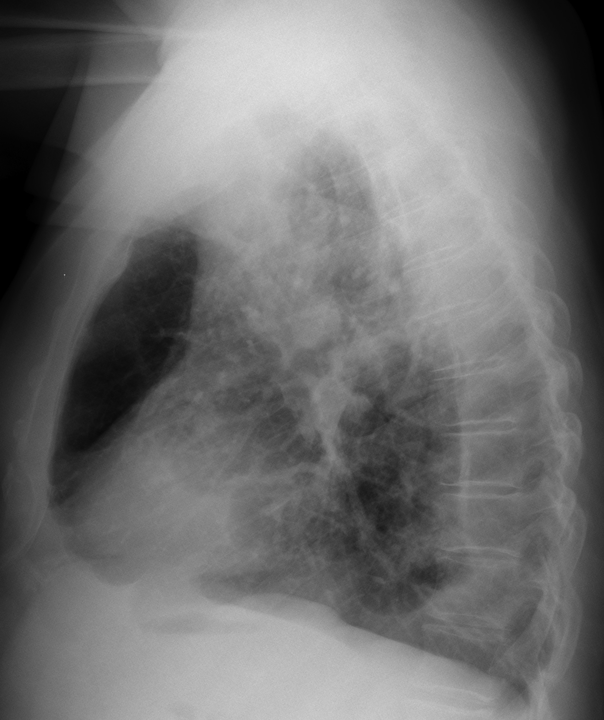
The chest radiograph (TB in youth) shows signs of chronic pleural disease: small hemithorax and calcified pleural thickening.
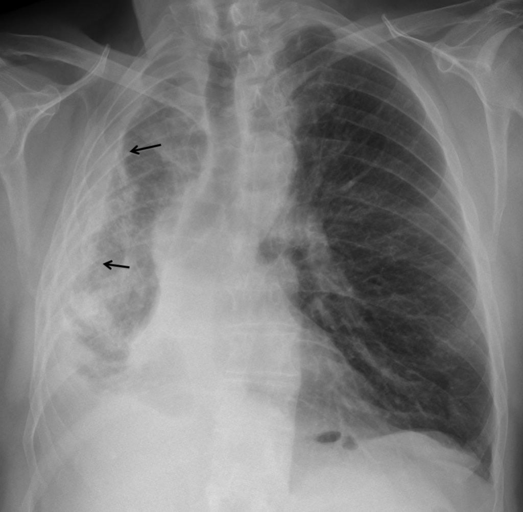
Fig. 1
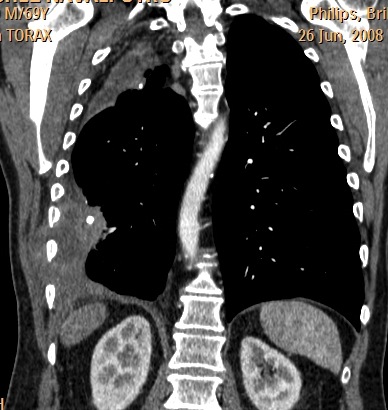
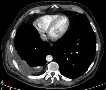
CT confirms the findings and shows a solid component (arrows) with invasion of the chest wall (arrows).
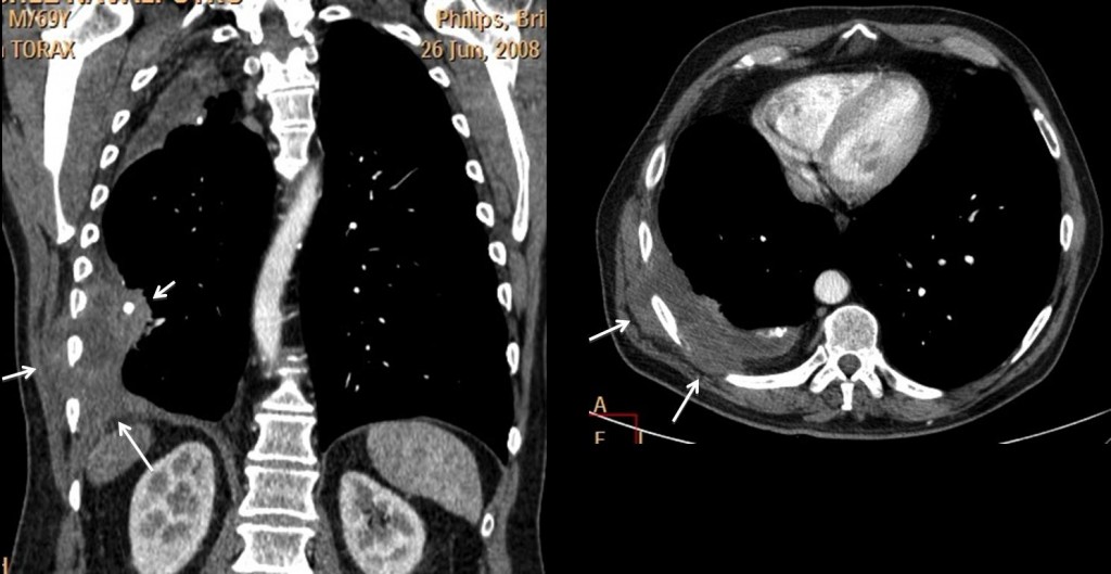
Fig. 2
This combination is highly suggestive of malignancy developing in chronic pleural disease (lymphoma, carcinoma or mesothelioma. They all look the same). Empyema necessitatis can be discarded because it is mainly fluid and has no solid component.
Final diagnosis: lymphoma developing in chronic pleural disease. Patient is alive and well four years after initial diagnosis. Ref. AJR 194:76-84; 2010
Teaching point: complications of chronic pleural disease are mainly three: bronchopleural fistula, empyema necessitatis and malignant tumour. CT can easily distinguish between the last two, because of the solid component of the tumour.