 Dear friends,
Since some of you have been complaining about the cases being difficult, Muppet wants to show a really difficult case: a 76-year-old woman with previous heart troubles and currently asymptomatic.
Diagnosis:
1. Calcified pericardial cyst
2. Hydatid cyst of the heart
3. Intracardiac calcified aneurysm
4. None of the above
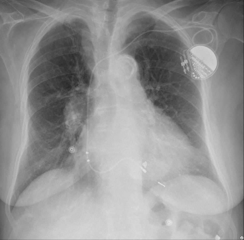
76-year-old- woman, PA chest
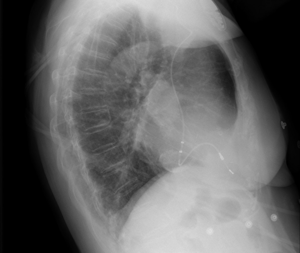
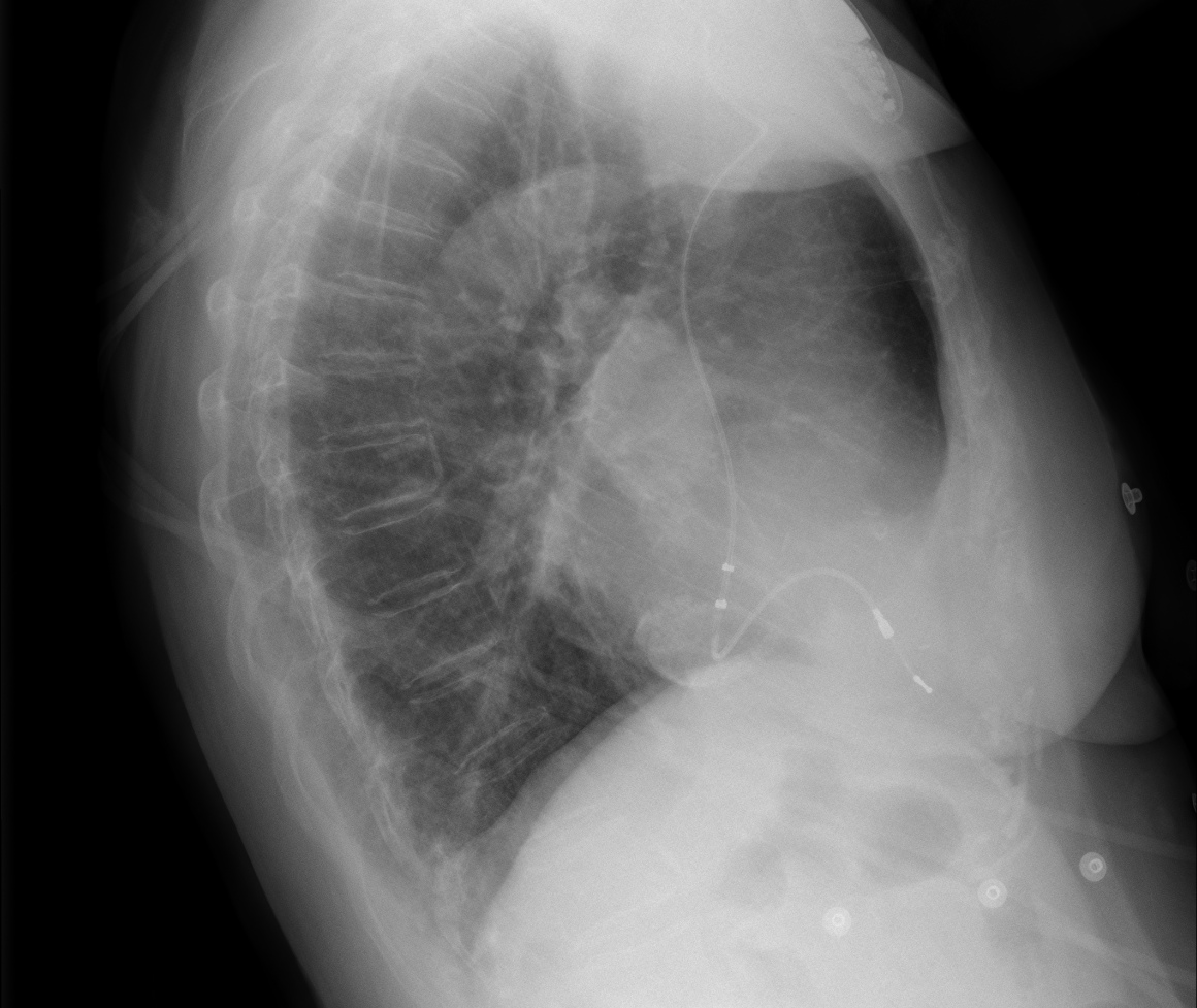
76-year-old- woman, lateral chest
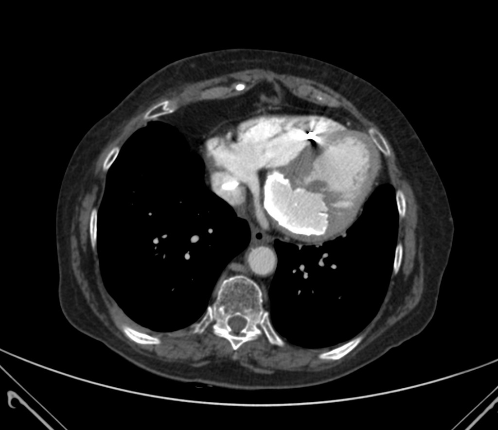
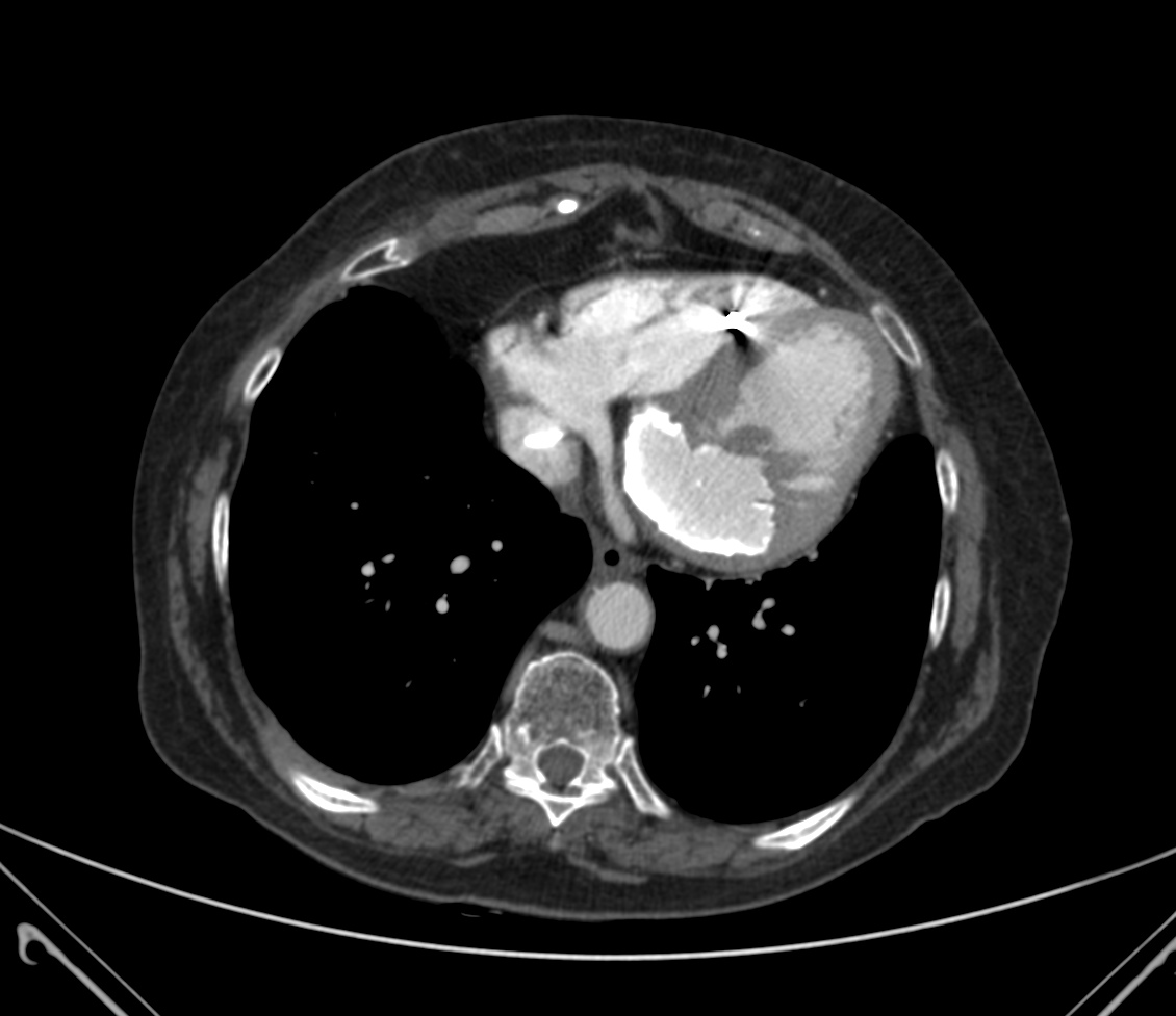
76-year-old- woman, axial CT
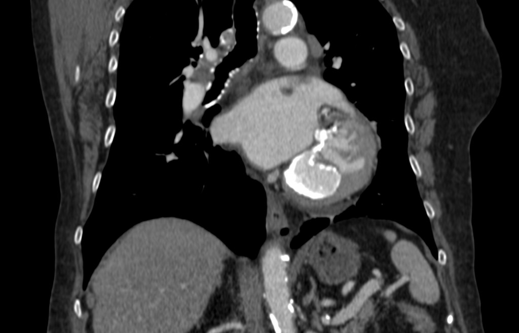
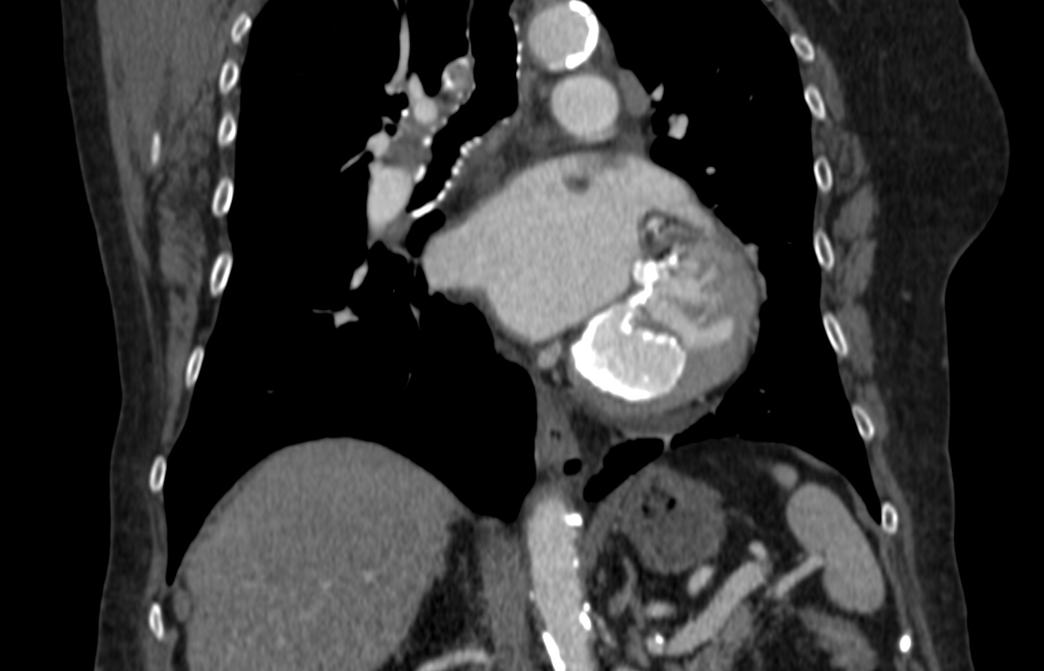
76-year-old- woman, coronal CT
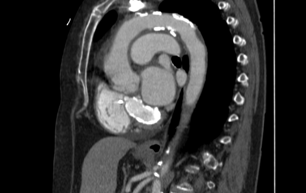
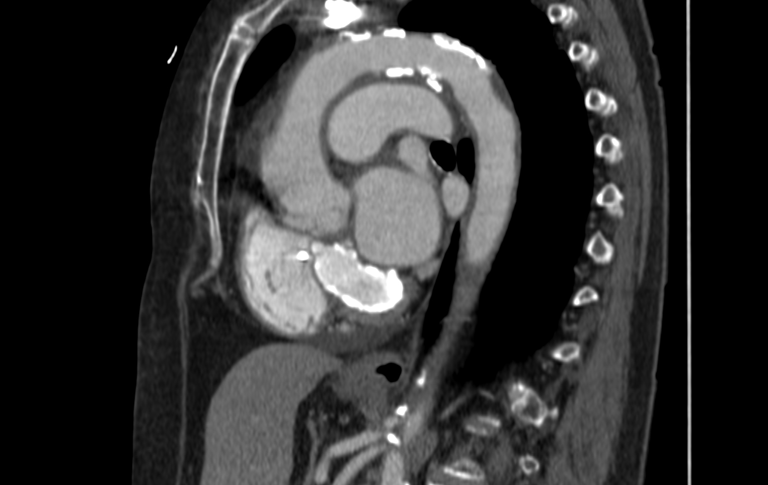
76-year-old- woman, sagittal CT
Click here for the answer to case #35
Since Muppet said this is a difficult case, most of you have made the correct diagnosis, just to spite him. The main finding is an oval calcified cardiac opacity, barely visible in the PA chest and better seen in the lateral view (arrows).
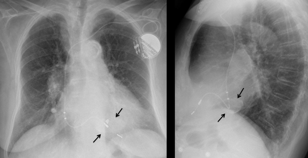
Fig. 1
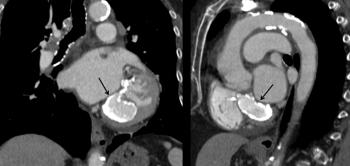
CT confirms the presence of the oval calcification (Fig. 2, arrows). Although the appearance is striking, the aspect is characteristic of caseous necrosis of the mitral annulus, an entity described in the eighties, due to degeneration of the annulus. It occurs in elderly patients and does not cause any symptoms. The word caseous is related to the central degenerative softening and has no relationship to tuberculosis.
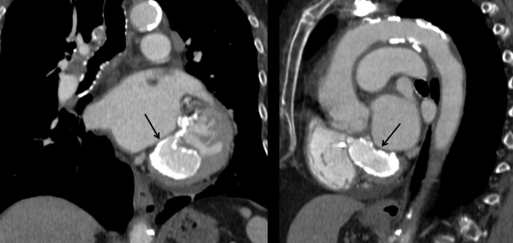
Fig. 2
Final diagnosis: caseous necrosis of mitral annulus.
Teaching point: it is important to recognise this entity to avoid confusing it with other pathologies. Muppet believes the plain film appearance is very typical. As far as he knows, it has not been described in the literature, because nobody cares about plain films anymore.
N.B. Some of you have complained that the images are too small. Muppet is happy to announce that he has been told a trick to enlarge them. Once the image is selected, hold down ‘control’ (cmd in Mac) and click the ‘+’ key. Every click increases the size of the image. To reverse, click ‘–’.