Dear Friends,
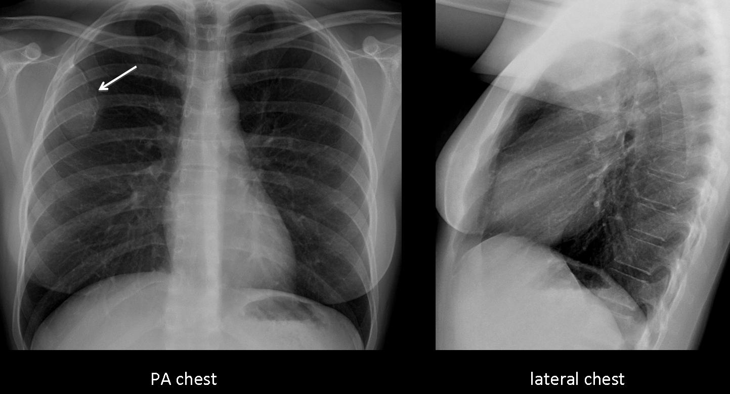
Since you are getting used to difficult cases, Muppet is showing you an easy one. May the Force be with you. It is a pre-op chest radiograph of a 37-year-old woman with breast carcinoma.
Diagnosis:
1. Metastases
2. Granuloma TB
3. Hydatid cyst
4. None of the above
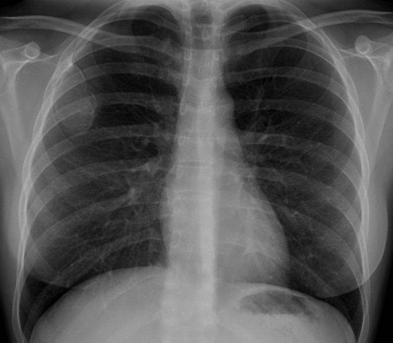
37-year-old woman, PA chest

37-year-old woman, lateral chest
Click here for the answer to case #36
Findings: the PA chest film shows a solitary expansive lytic lesion in the anterior aspect of the third right rib (arrow), without an accompanying soft tissue mass.
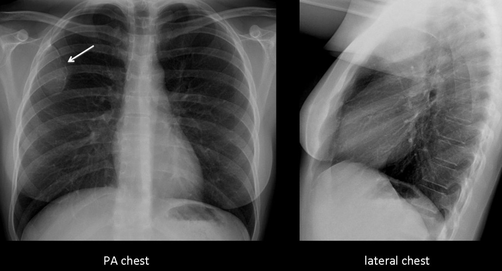
Fig. 1
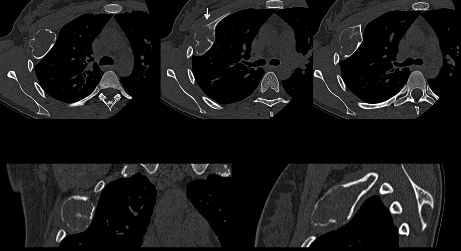
CT confirms the findings and the lack of soft tissue component. There is cortical thinning and a short transition zone with normal bone (arrow). The appearance of the lesion is very suggestive of a benign process and, as many of you pointed out, fibrous dysplasia is the first possibility.
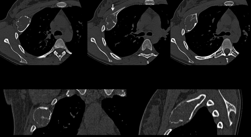
Fig. 2
Final diagnosis: fibrous dysplasia.
Teaching point: Solitary expansive lesion in a rib in a young person is highly indicative of fibrous dysplasia, which is the most common benign lesion of ribs.