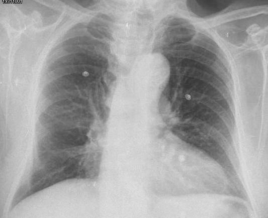
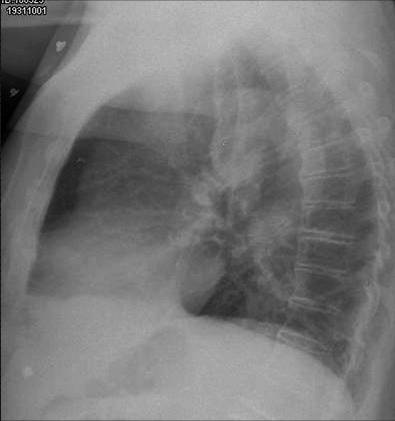
Muppet is impressed with your knowledge. He tries very hard to post teaching cases and has decided to skip the multiple-choice questions in the following chest case, which is that of an asymptomatic 56-year-old male.
1. Where is the lesion?
2. What is your diagnosis?
The first five radiologists to suggest the correct diagnosis will be given a DVD at the next European Congress of Radiology.
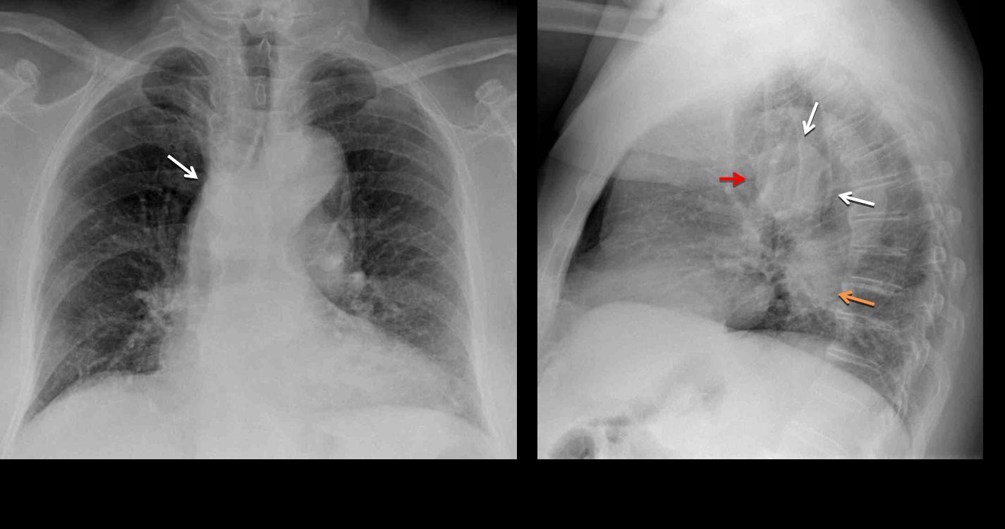
Chest radiographs show a rounded opacity in the middle mediastinum (white arrows), imprinting in the lower trachea (
red arrow). There is a lower rounded opacity (
orange arrow), which represents the tortuous descending aorta.
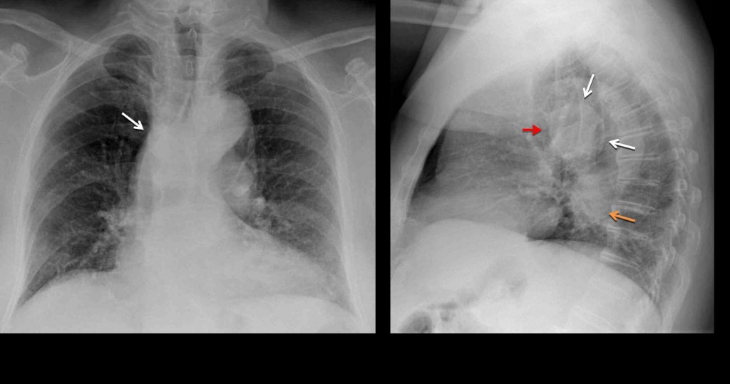
Fig. 1
Answering the first question, the abnormality is located in the middle third of the middle mediastinum. Most common masses in this location are lymph nodes and congenital duplication cysts. However, the location in the PA view and the imprint in the lower trachea should suggest an aberrant left pulmonary artery (pulmonary sling), as Raffaela mentioned.
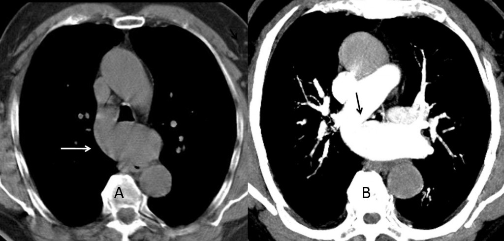
Fig. 2
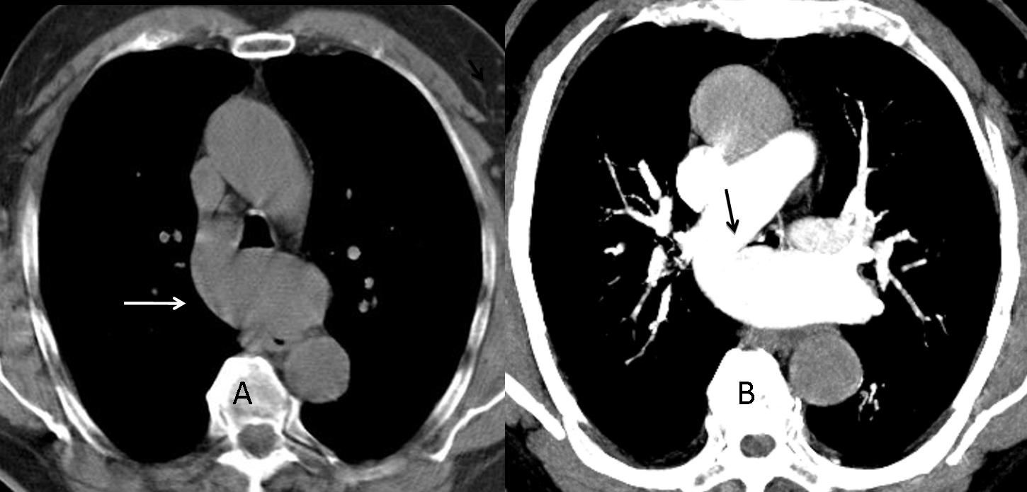
CT confirms the diagnosis by showing the anomalous artery (A, arrow) crossing between the oesophagus and trachea and the origin of the left pulmonary artery from the right (B, arrow).
Diagnosis: aberrant left pulmonary artery (pulmonary sling).
Congratulations to Raffaela for her excellent diagnosis. You can choose from the Godfather trilogy, The Lord of the Rings, or The Bourne trilogy.
Teaching point: always look at the trachea in the lateral film. It is an excellent marker for discovering middle mediastinal masses.