Muppet is getting old and soft and from now on will show only easy cases. Today’s case is a 33-year-old girl with fever and malaise.
1. Lymphoma
2. TB
3. Wegener’s
4. None of the above
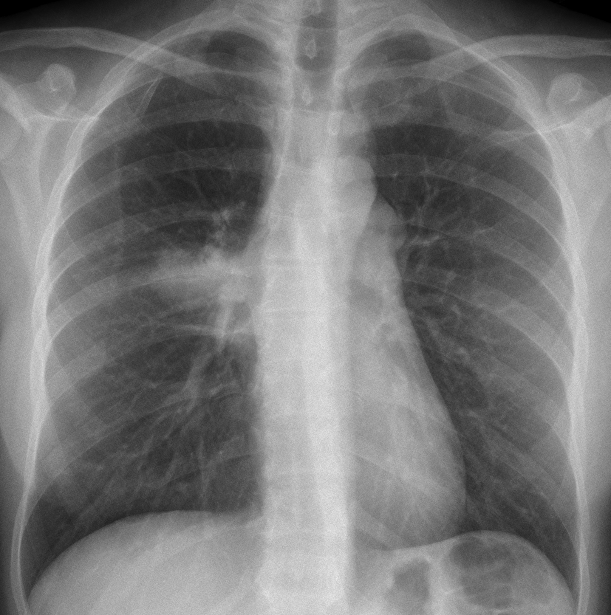
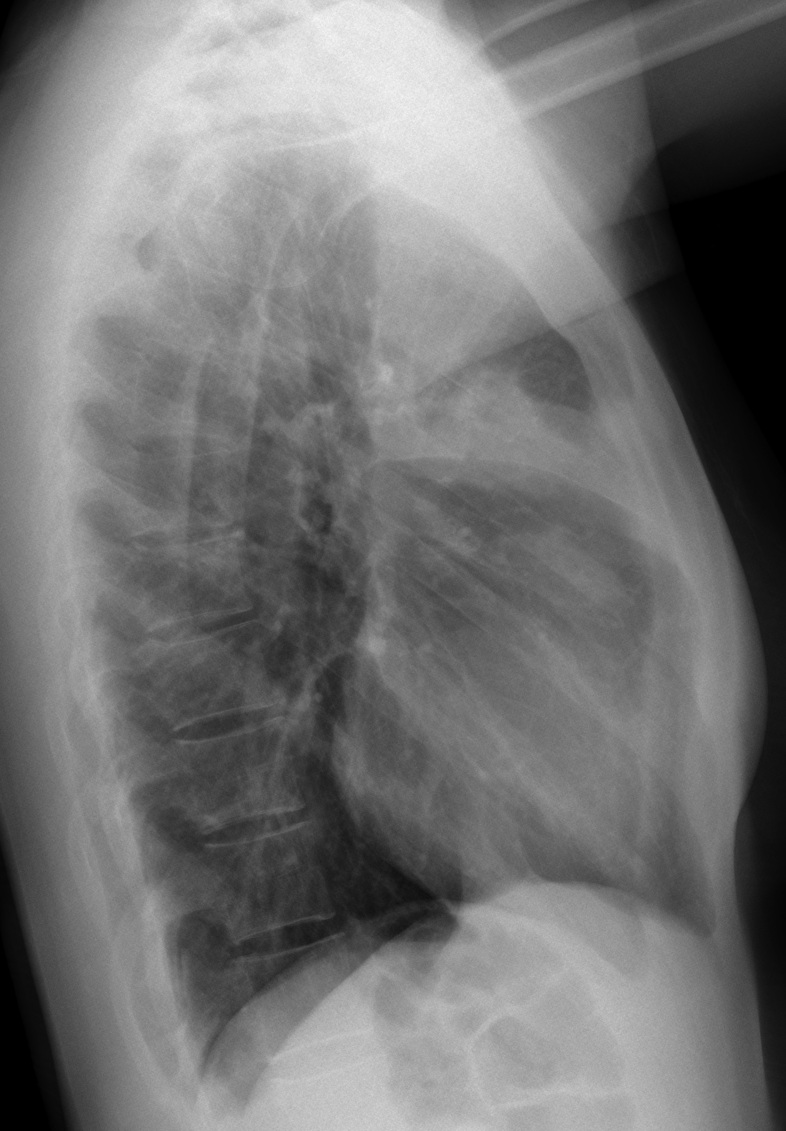
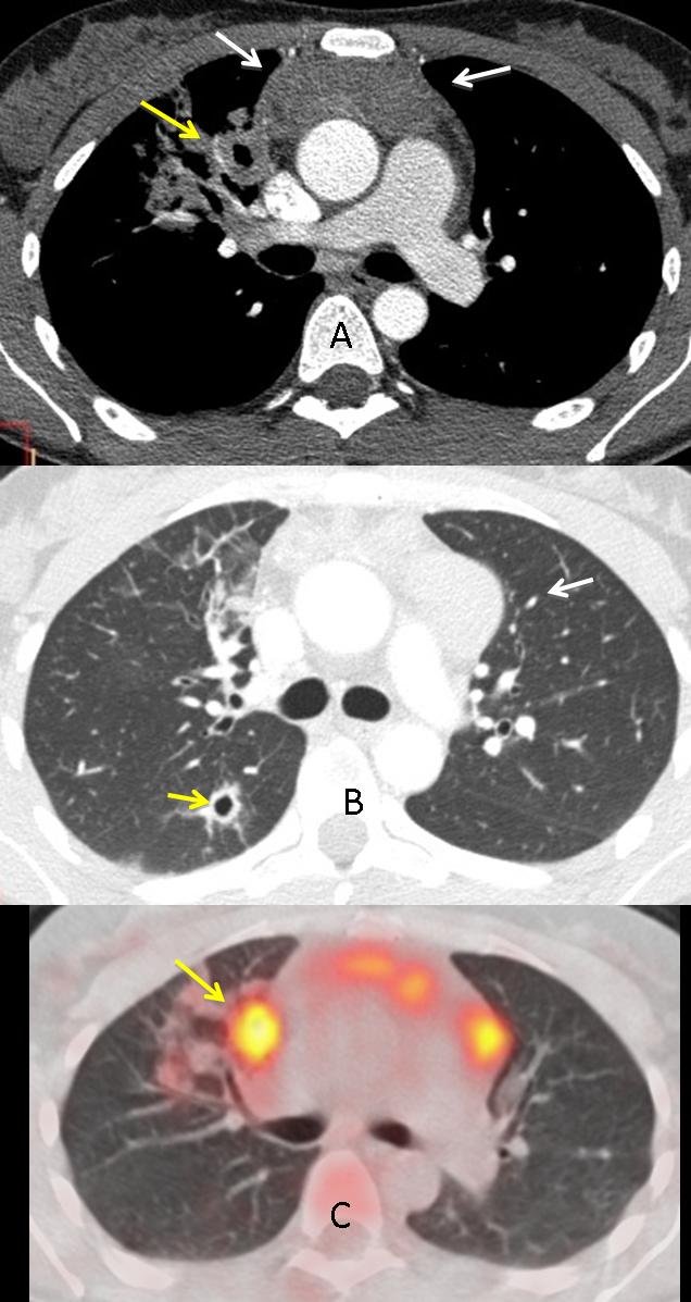
Findings: chest radiographs show a parahilar right pulmonary opacity located in the anterior segment of RUL in the lateral view (white arrows). There are lymph nodes in the aorto-pulmonary window (red arrow).
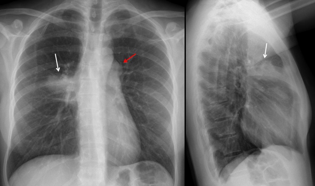
Fig. 1
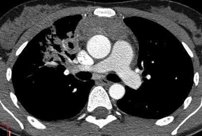
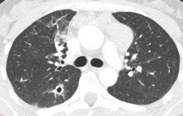
CT shows anterior mediastinal lymphadenopathy (white arrows), cavitating pulmonary lesions (A, B, yellow arrows) and a right pulmonary infiltrate. PET-CT shows uptake of lymph nodes and pulmonary nodule (C, arrow).
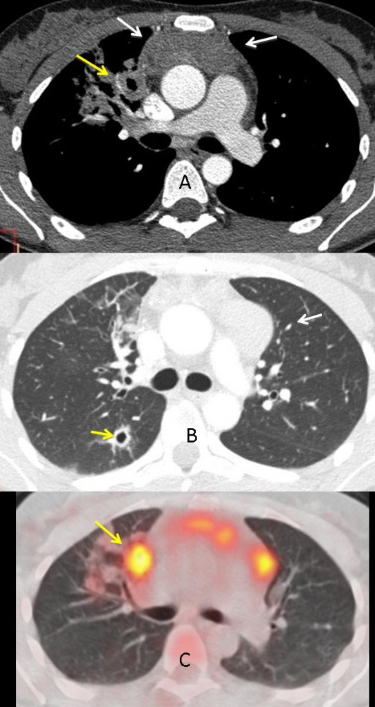
Fig. 2
Of the diagnoses suggested, Wegener’s is unlikely; lymphadenopathy is uncommon; and the pulmonary infiltration and nodules are confined to a single area. TB is a possibility, but the presence of non-necrotic lymph nodes in the anterior mediastinum, with none in the hilum, goes against it.
Lymphoma is the most likely diagnosis: usually present as anterior mediastinal lymphadenopathy, may invade pulmonary parenchyma by contiguity and occasionally is associated with cavitating pulmonary nodules.
Final diagnosis: Hodgkin’s disease, nodular sclerosis type.
Teaching point: when confronted with multiple findings, try to determine the most valuable. In this case, the anterior mediastinal lymphadenopathy, which suggests lymphoma in a young symptomatic patient.