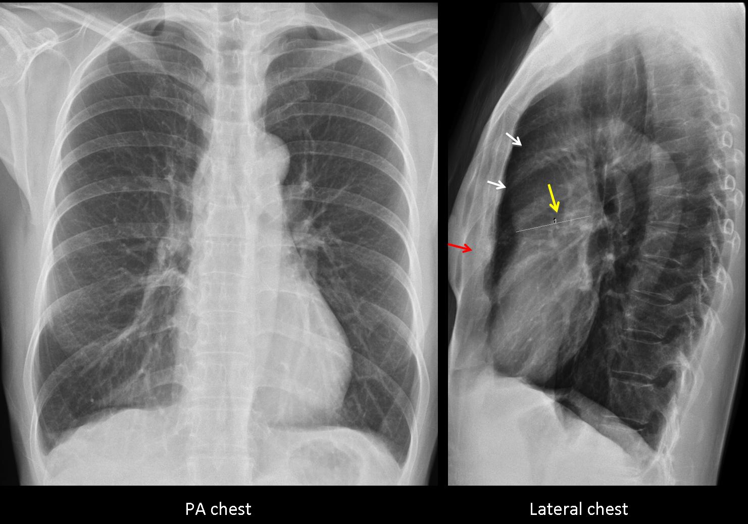
Since we had very few answers in the previous case (too easy or too difficult?), Muppet is reverting to plain films and straightforward questions. The following are pre-employment radiographs of a 43-year-old female. She was told that she had enlargement of the ascending aorta. What would be your diagnosis?
1. Marfan’s
2. Aortic coarctation
3. Aortic valvular disease.
4. None of the above
Findings: PA chest film is unremarkable. The ascending aorta looks normal. Lateral film shows a prominent ascending aorta (white arrows). A line has been traced which measures an apparent dilatation of the ascending aorta (yellow arrow). The obvious error is that the line is drawn from the anterior aspect of the aorta, outlined by lung air, to an imaginary point in the mediastinum, which may or may not be the posterior aortic wall. It is important to remember that the inner wall of the aorta contacts with the mediastinal tissues and, according to the silhouette sign, cannot be visible. Therefore, whoever traced the line is wrong.
MRI confirmed that the aorta was normal and that no other abnormalities were present. The apparent prominence of the ascending aorta in the lateral film is due to a combination of a moderate pectus (red arrow) and a narrow AP diameter of the chest.
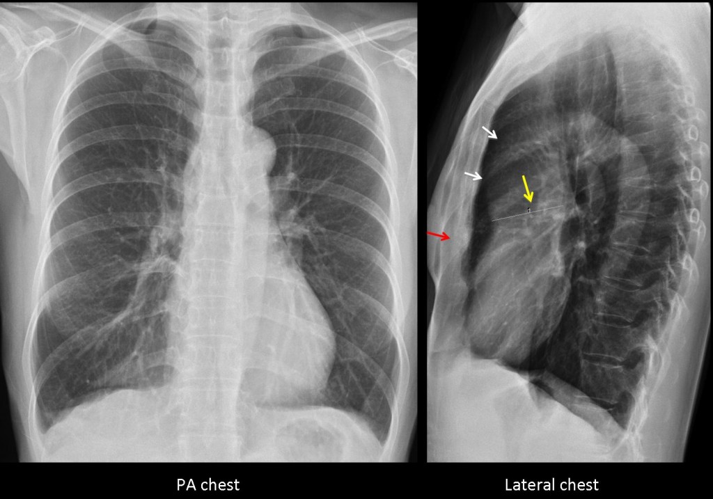
Fig. 1
The patient is friend of mine and the examination was made in a university hospital abroad. MRI is not shown because I could not obtain it.
Final diagnosis: normal chest
Teaching point: do not forget that in the chest radiograph we only see the outer contour of the aorta. It is impossible to see the inner wall because it blends with mediastinal structures. Remembering this basic principle will avoid errors like the one that happened in this patient.
And never, never, draw lines in the radiographs!