Dear friends,
Welcome back to Caceres’ corner.
I’m already keen on your suggestions on Case #4.
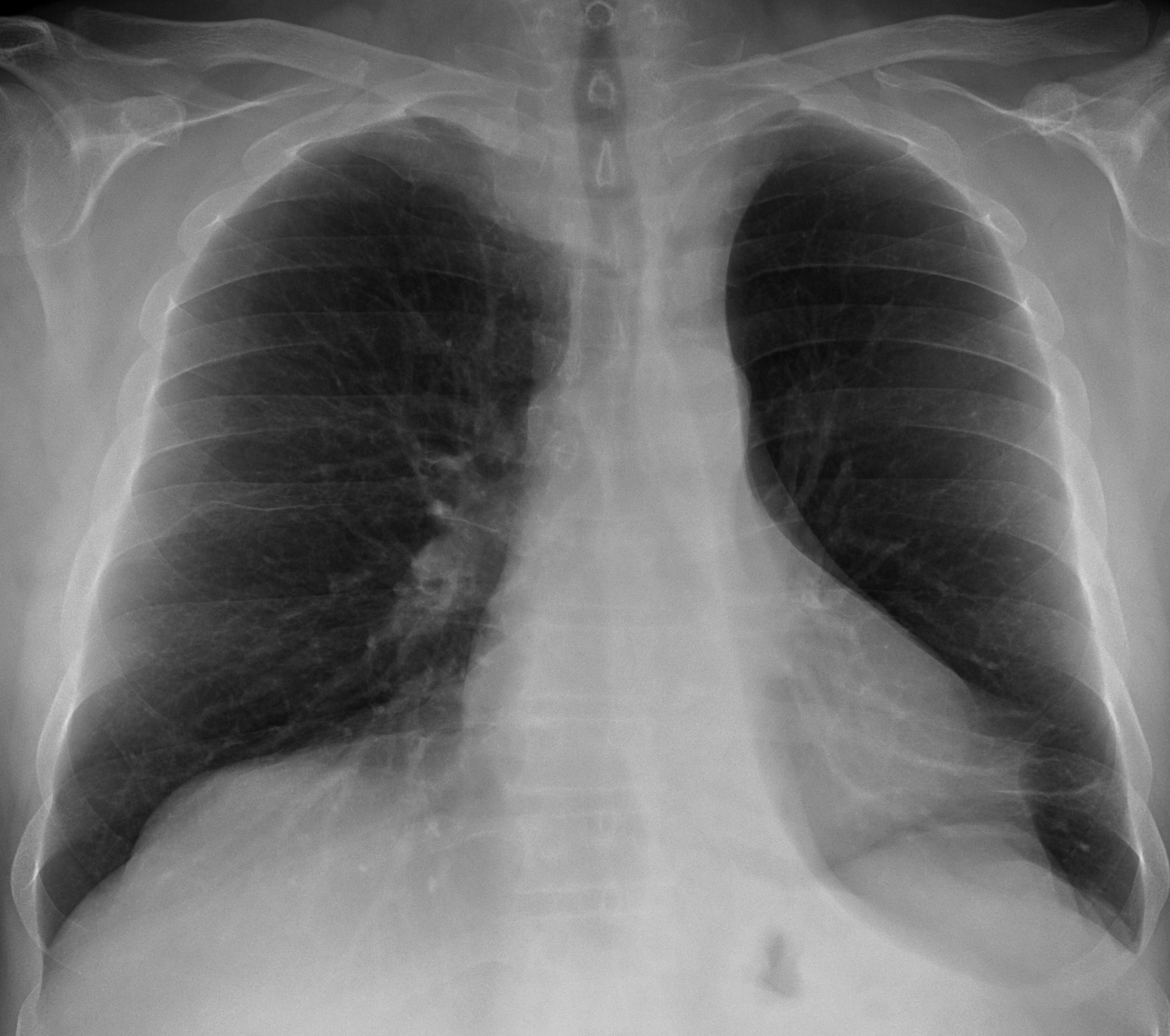
Clinical history: Pre-operative chest radiograph for ophthalmologic surgery in a 57 y. o. male
Most likely diagnosis:
1. Carcinoma of the lung
2. Unilateral hyperlucent lung
3. Pulmonary embolism
4. Giant bulla
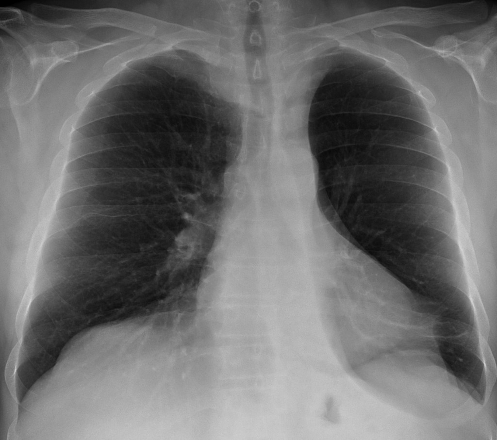
Pre-operative chest radiograph for ophthalmologic surgery in a 57 y. o. male (PA chest)
Click here for the answer to case #4
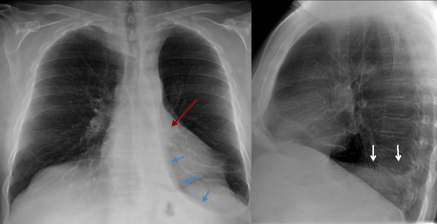
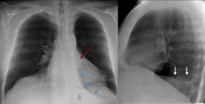
My initial impression of the PA chest is a left hyperlucent lung with diminished vascularity. Muppet is smarter and always looks for other findings. He sees a very low left hilum (red arrow), a paraspinal oblique line (blue arrows) and blurring of the left hemidiaphragm on the lateral view (white arrows). These findings are very suggestive of left lower lobe collapse. The most common causes of collapse of the lower lobes are bronchiectasis and carcinoma. Considering that the former was not included in the options, the most likely diagnosis is carcinoma of the lung.
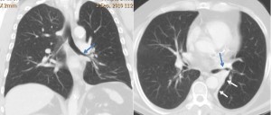
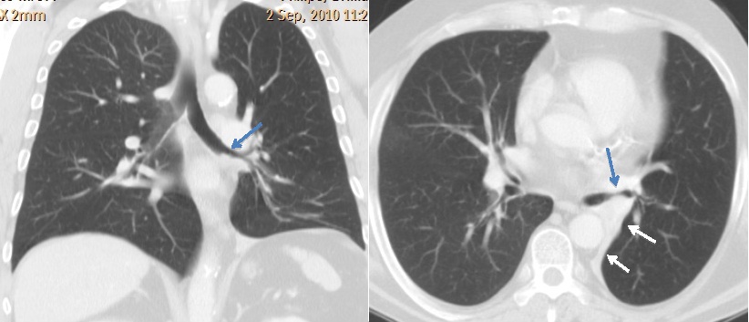
Diagnosis is confirmed with CT, which shows irregular narrowing of the bronchus (blue arrows) and marked collapse of LLL (white arrows). Biopsy: epidermoid carcinoma.
Teaching point: remember case #1. Always look at the hila. They are very useful markers in lobar collapse. Muppet will be very disappointed if you do not remember this!