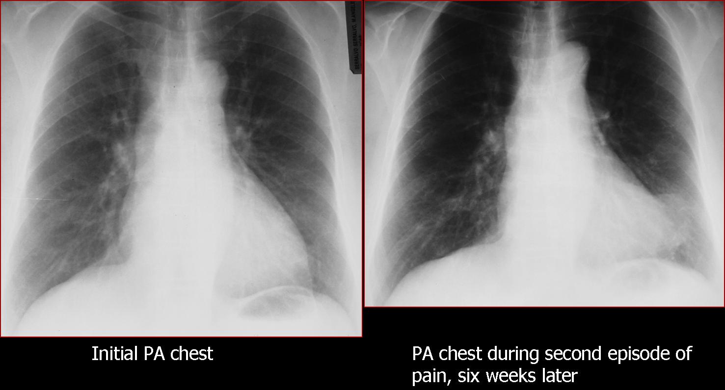
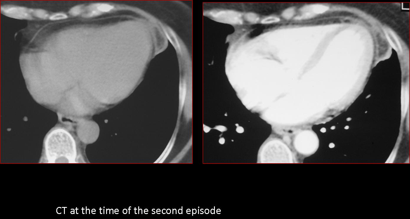
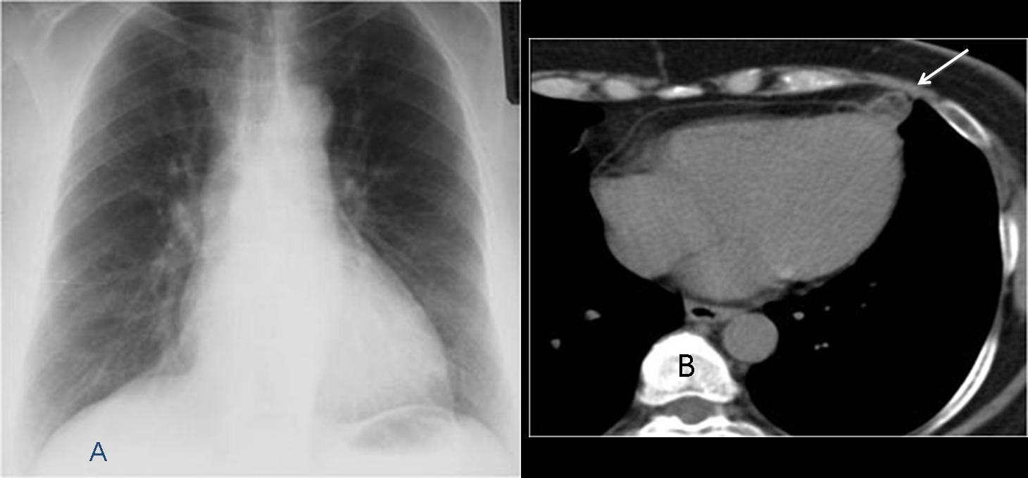
Muppet is so happy with your performance that he has chosen a challenging case. The following radiographs belong to a 52-year-old woman with two episodes of chest pain in six weeks. The initial radiograph is shown, as well as a radiograph taken during the second episode. At that time, a CT was done.
1. Pulmonary infarct
2. Lymphadenopathy
3. Loculated pericardial fluid
4. None of the above
Answer to case 40
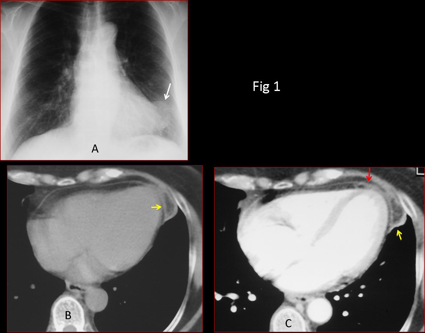
Initial PA chest shows a paracardiac mass (Fig 1A, arrow). In CT the mass is of fatty density, with a capsule that enhances after injection of IV contrast (B,C yellow arrows). There is mild pericardial thickening (C, red arrow).
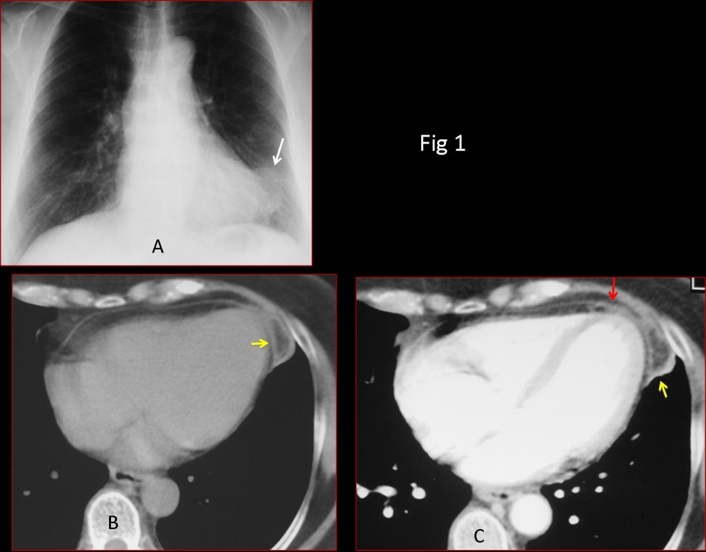
Fig. 1
The appearance is pathognomonic of epipericardial fat necrosis. Diagnosis is confirmed with follow-up imaging. The mass has shrunken markedly on CT (Fig 2B, arrow) and is no longer visible in the chest radiograph.
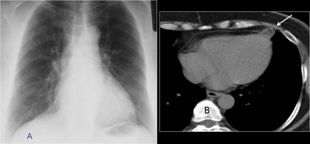
Fig. 2: Two months later
Final diagnosis: epipericardial fat necrosis.
Congratulations to Muppet’s faithful followers, who attained 100% correct answers (didn’t expect any less of you!). They may not know that this entity was described in the radiological literature in 2005. The initial description was made in 1957 and only 20 cases had been reported since; all of them operated on. Since 2005, this entity is being described with increased frequency (aside from isolated case reports, two educational exhibits at the ARRS meetings in 2011 and 2012 showed 10 and 14 cases respectively).
Teaching point: EFN is an easily recognisable entity, more common that believed and should be considered in the differential diagnosis of acute chest pain.