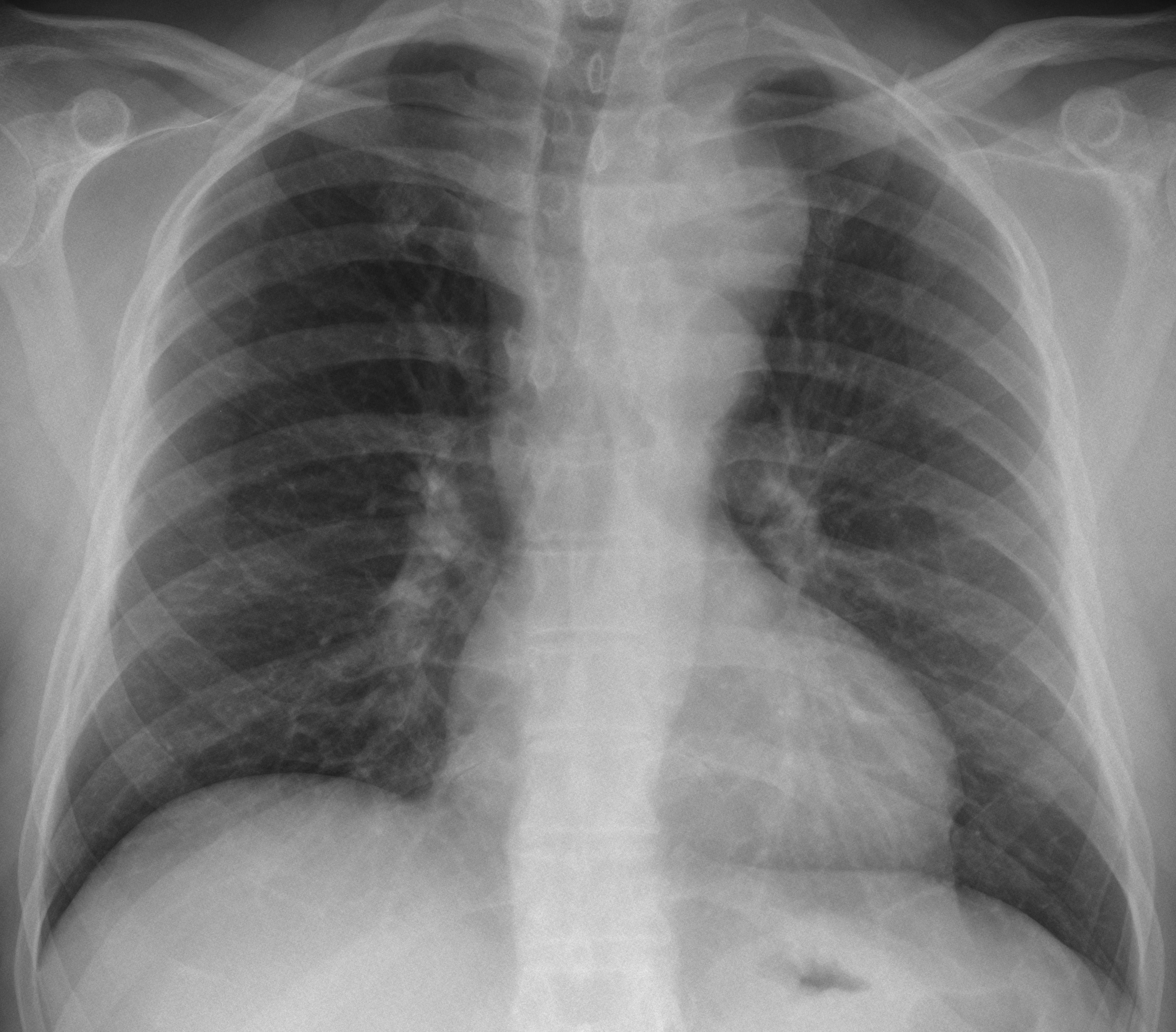
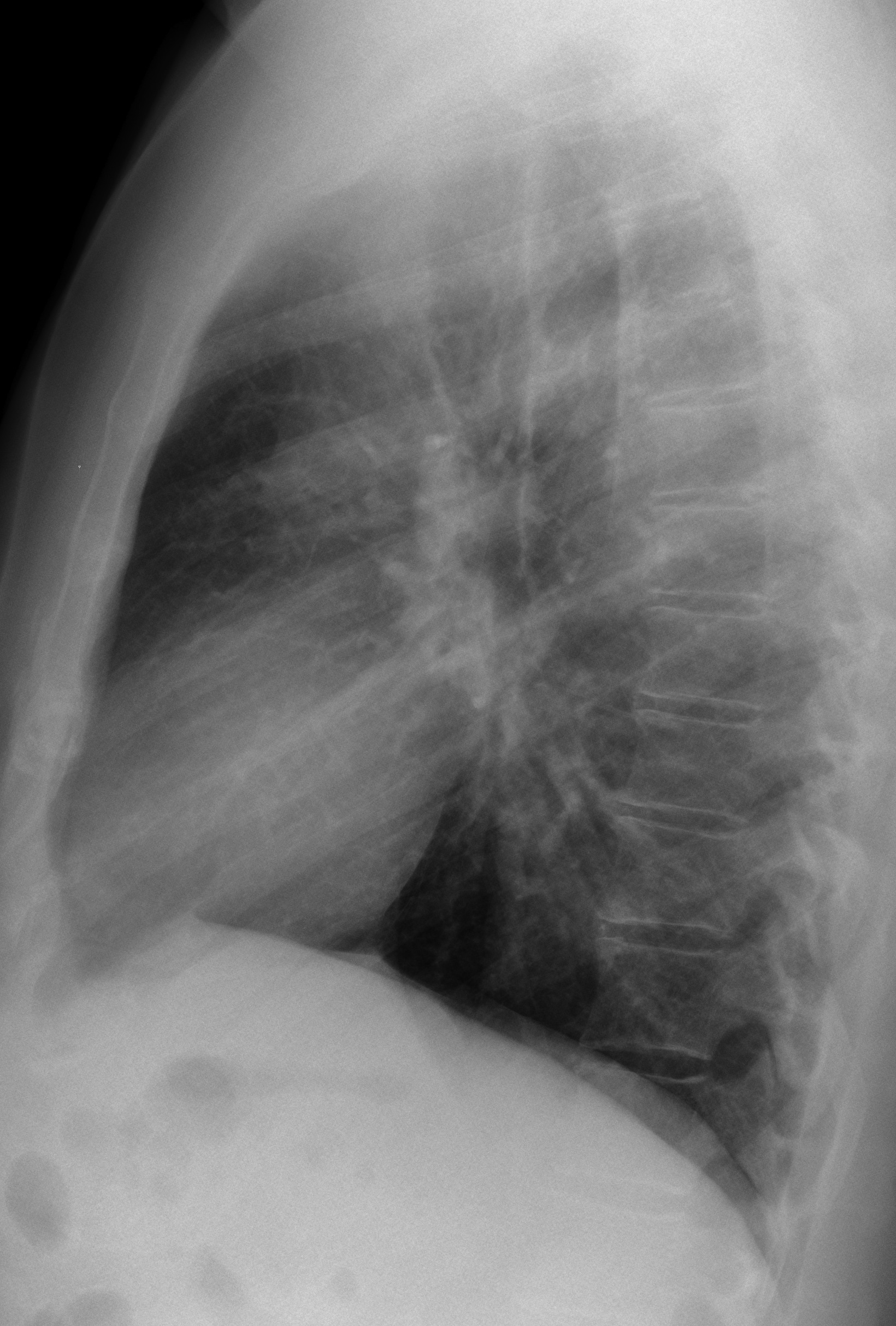
Muppet is seriously considering retirement, seeing your level of expertise! You have nailed the previous two cases. We hope you diagnose the following one just as well: 45-year-old man with vague chest pains.
1. Aortic aneurysm
2. Intrathoracic goiter
3. Thymoma
4. None of the above
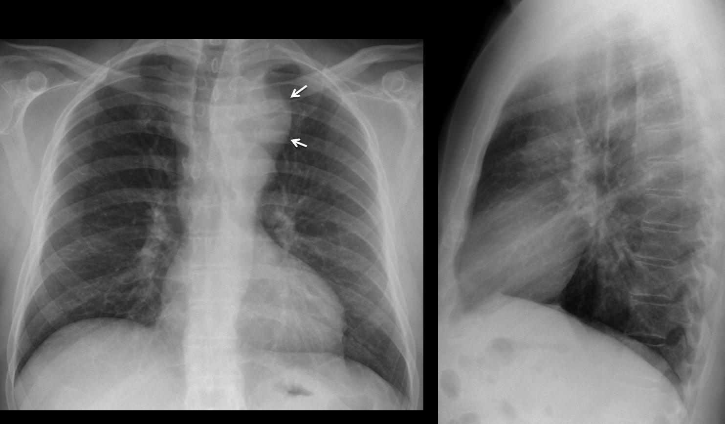
Findings: PA chest radiograph shows a superior middle mediastinal mass (Fig. 1, arrows), displacing the trachea. Mass is not well seen on the lateral view. The most common diagnosis should be an endothoracic goiter, but remember that a vascular structure should always be ruled out with enhanced CT.
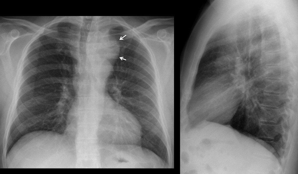
Fig. 1
Coronal CT demonstrates that the mass corresponds to a dilated aortic arch (arrow), located higher than usual (Fig. 2A, arrow). On the oblique reconstruction, the high aortic arch is easily seen, as well as an indentation (Fig. 2B, arrow) at the junction of the aneurysmatic arch and the descending aorta.
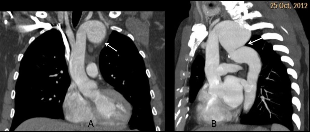
Fig. 2
Final diagnosis: aortic pseudocoarctation with aneurysm of the aortic arch.
Pseudocoarctation of the aorta occurs when the aortic arch originates from the 3rd arch, instead of the 4th. In this condition, the aortic arch is higher than usual, with a kinking at the union of the aortic arch and descending aorta, simulating aortic coarctation. Rib notching is absent and systemic hypertension is not present. This condition is unusual and I have seen several cases, but none with aneurysm, which was discovered when seeing this patient a few weeks ago. This demonstrates that we should always be ready for the unexpected!
Congratulations to Dr. Froso, who gave an excellent discussion.
Teaching point: remember that the mediastinum is composed basically by vessels. When seeing a mediastinal mass on the plain film always perform an enhanced CT to rule out a vascular origin.