Dear Friends,
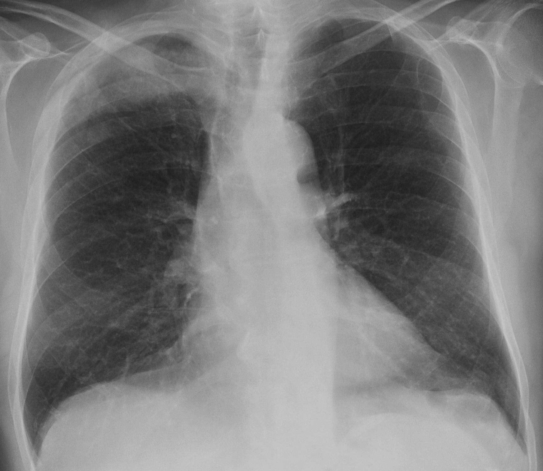
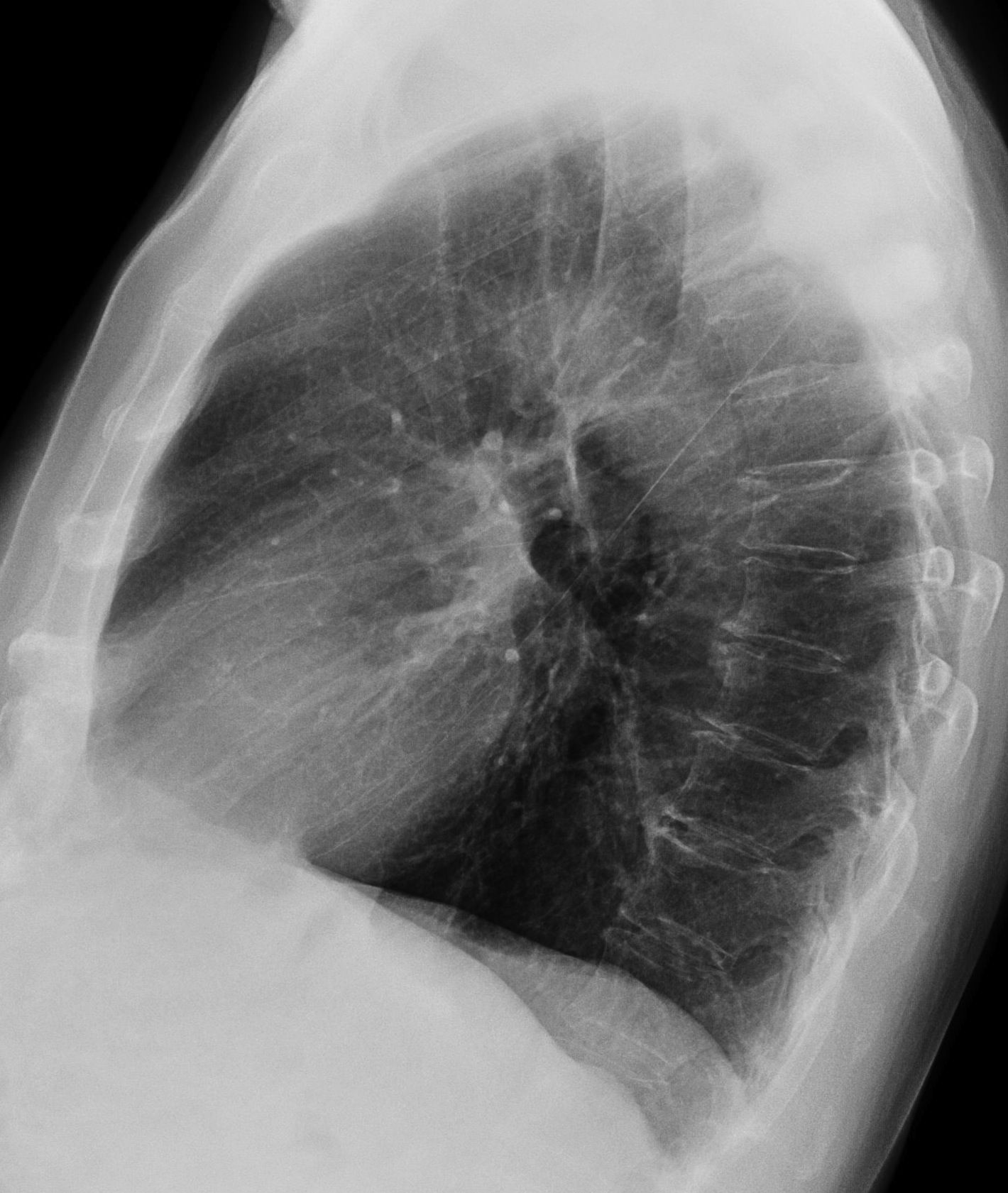
After the trepidation of case 42, Muppet hopes you get this case easily. Sixty-eight-year-old male with pain in the chest.
Diagnosis:
1. Tuberculosis
2. Bronchoalveolar carcinoma
3. Actinomycosis
4. None of the above
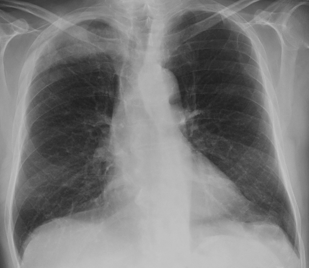
68-year-old male, PA chest
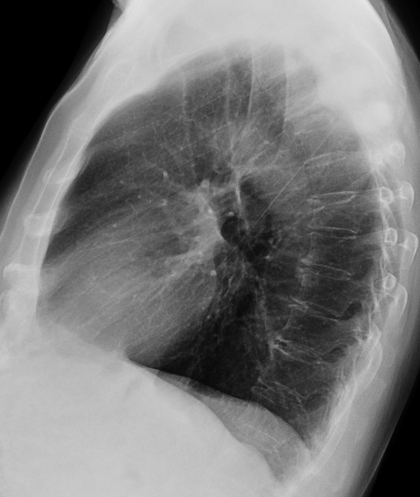
68-year-old male, lateral chest
Post your thoughts and diagnosis below and check back next Tuesday for the answer.
Click here for the answer to Case 43
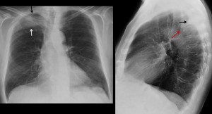
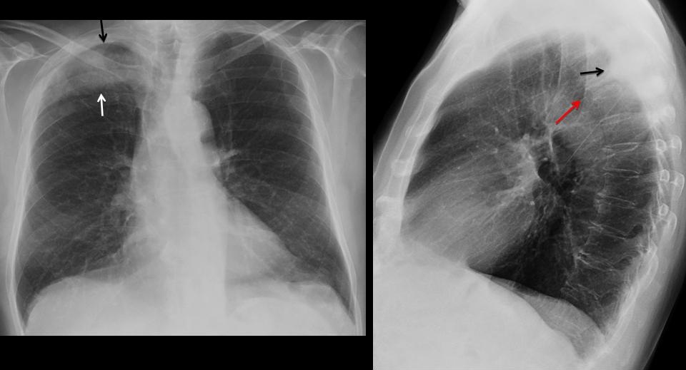
Findings: PA chest radiograph shows an ill-defined opacity in the right apex (arrows). Careful examination shows that the third rib is missing. On the lateral view there is a hint of an extrapulmonary lesion (arrow) and collapse of one of the upper vertebrae (red arrow).
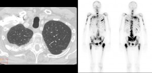
CT confirms the rib lesion, with a significant soft-tissue mass (arrows), which creates the illusion of a pulmonary infiltrate in the chest radiograph. Bone scan shows multiple areas of bone involvement.
Final diagnosis: bone metastases from prostate carcinoma.
From RSNA 2012 in Chicago, Muppet congratulates all of you who made the right diagnosis, especially Dr. Genchi Bari, who made some astute suggestions leading to the correct diagnosis
Teaching point: rib lesions may occasionally simulate pulmonary infiltrates. Always look at the ribs and you may be rewarded.