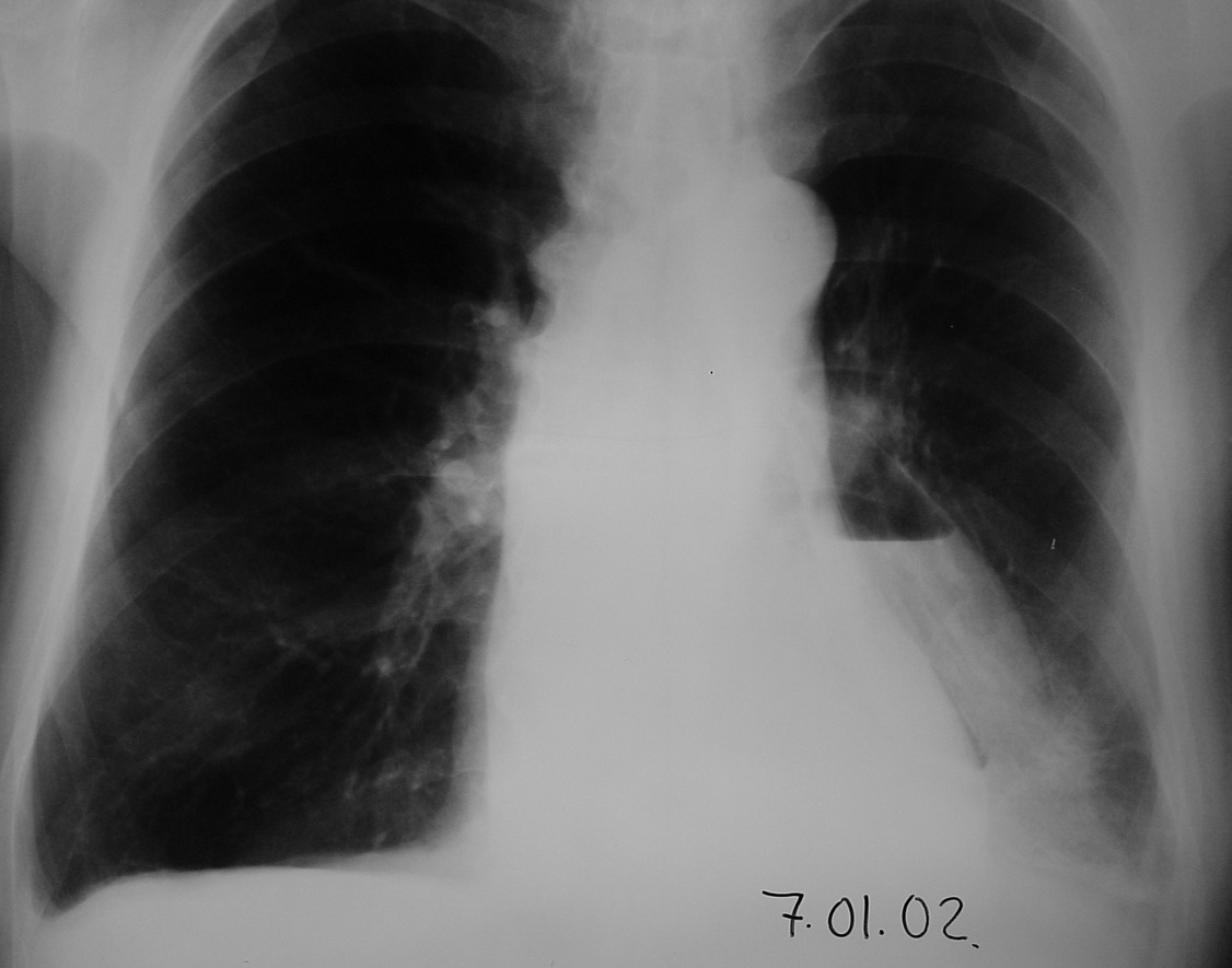
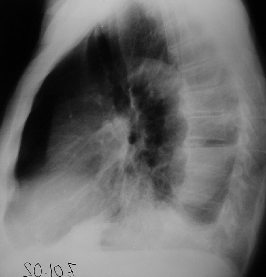
We are having a December sale (everything must go!) and during this month Muppet will show only cases with air-fluid level. We start with a vintage case (2002) of a 76-year-old male with left lower lobe pneumonia.
1. Infected bulla
2. Congenital cyst
3. Pneumatocele
4. None of the above
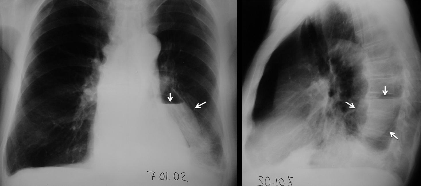
Findings: PA chest shows an air-space opacity in the left lower lobe, compatible with the clinical diagnosis of pneumonia. In addition, there is an elongated thin-walled cavity with an air-fluid level (arrows), located posteriorly in the lateral view (arrows).
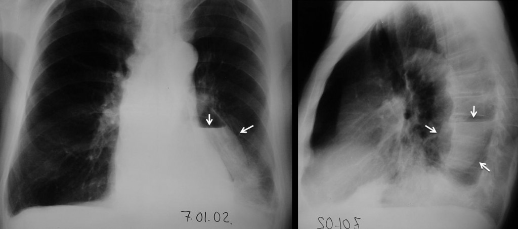
Fig. 1
Follow-up films show that the cavity is diminishing in size. The air-fluid level is now clearly within the left major fissure (Fig. 2a arrows). Later film shows that the air has disappeared, with a small amount of fluid remaining (Fig. 2b arrow).
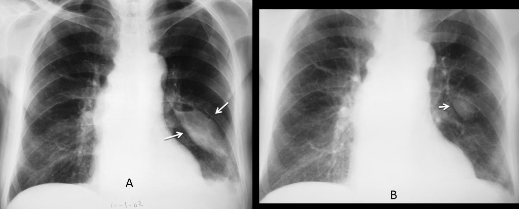
Fig. 2
Final diagnosis: loculated hidropneumothorax in the major fissure.
Muppet intends this case to be his Christmas present, because it is a rare condition and you probably did not know about it. Muppet is ashamed to confess that he does not know the exact etiology of this finding, although he suspects it is due to air seeping through the fissure layers.
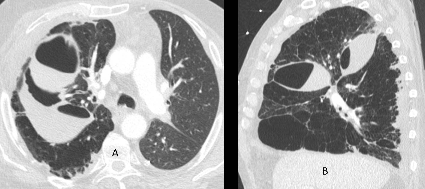
Fig. 3 shows another recent case, secondary to lung transplant, with air in minor and major fissures (arrows).
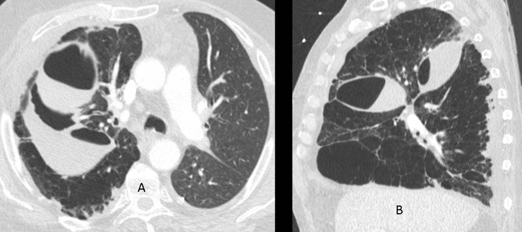
Fig. 3
Congratulations to Dr. Strangelove. He was the only one to suggest the diagnosis.
Teaching point: there is no obvious teaching point in this case. It is just an unusual image, easily recognisable if you think of it, and that should be included in the differential diagnosis of thin-wall cavities.