Dear Friends,
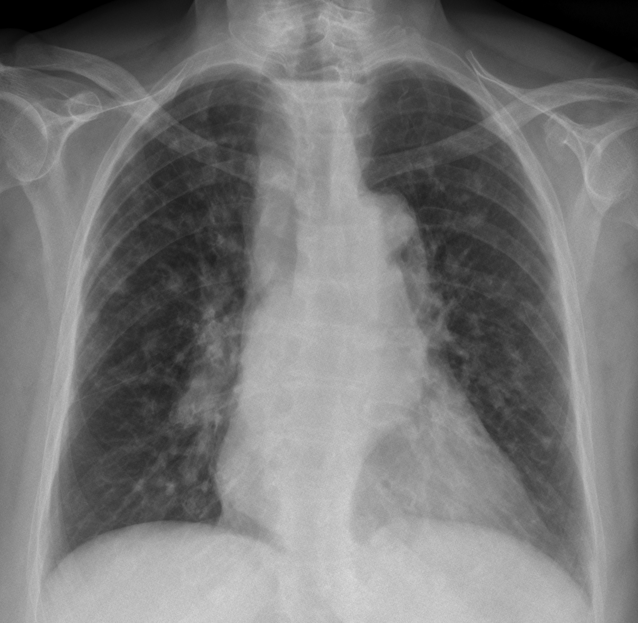
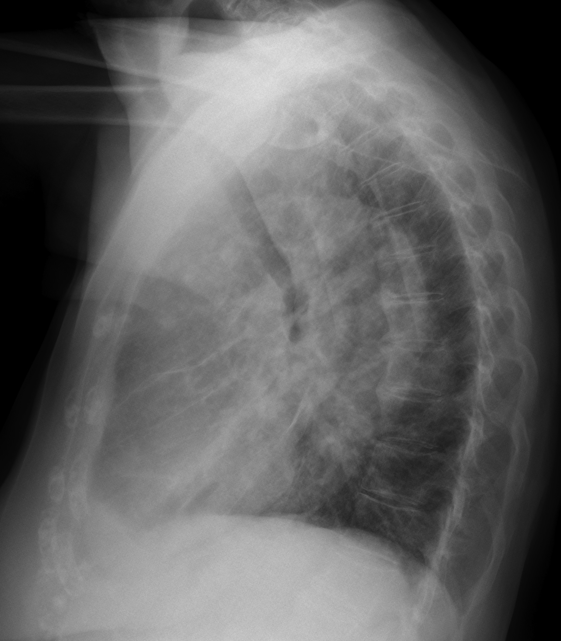
In the best Christmas spirit, Muppet is presenting a nice case. These radiographs belong to an 86-year-old lady who came to the emergency room with chest pain. Pulmonary nodules were visible. What would be your diagnosis?
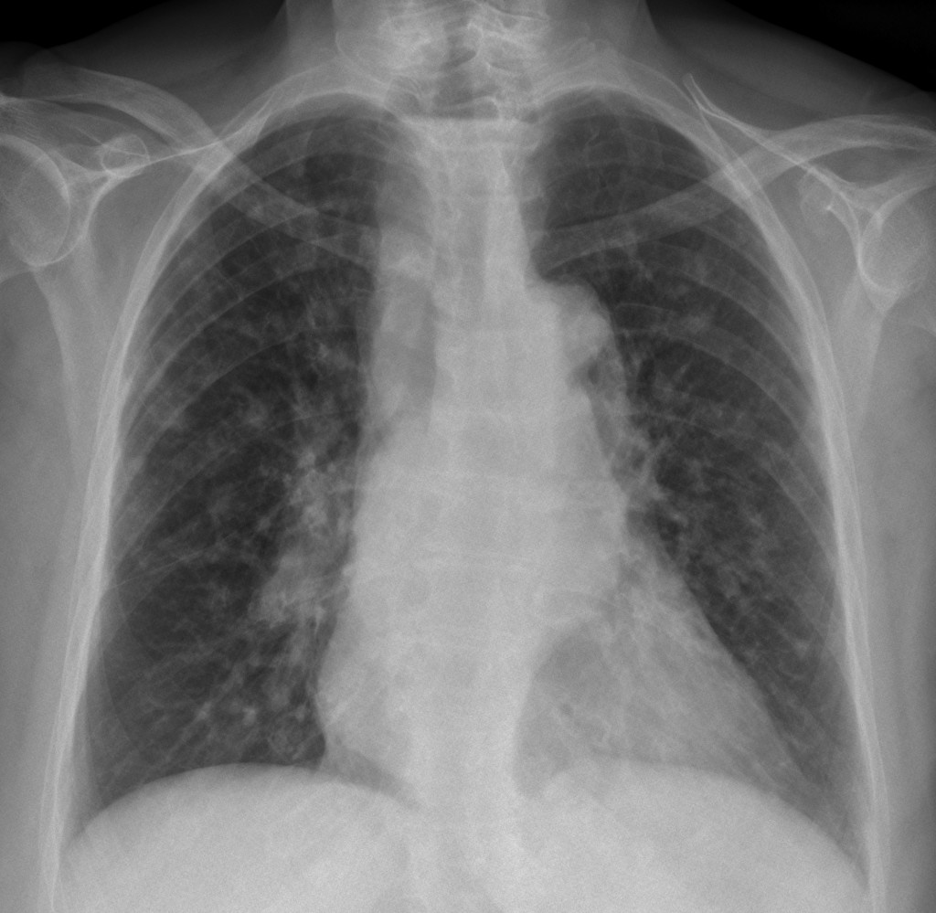
PA chest, 86-year-old woman
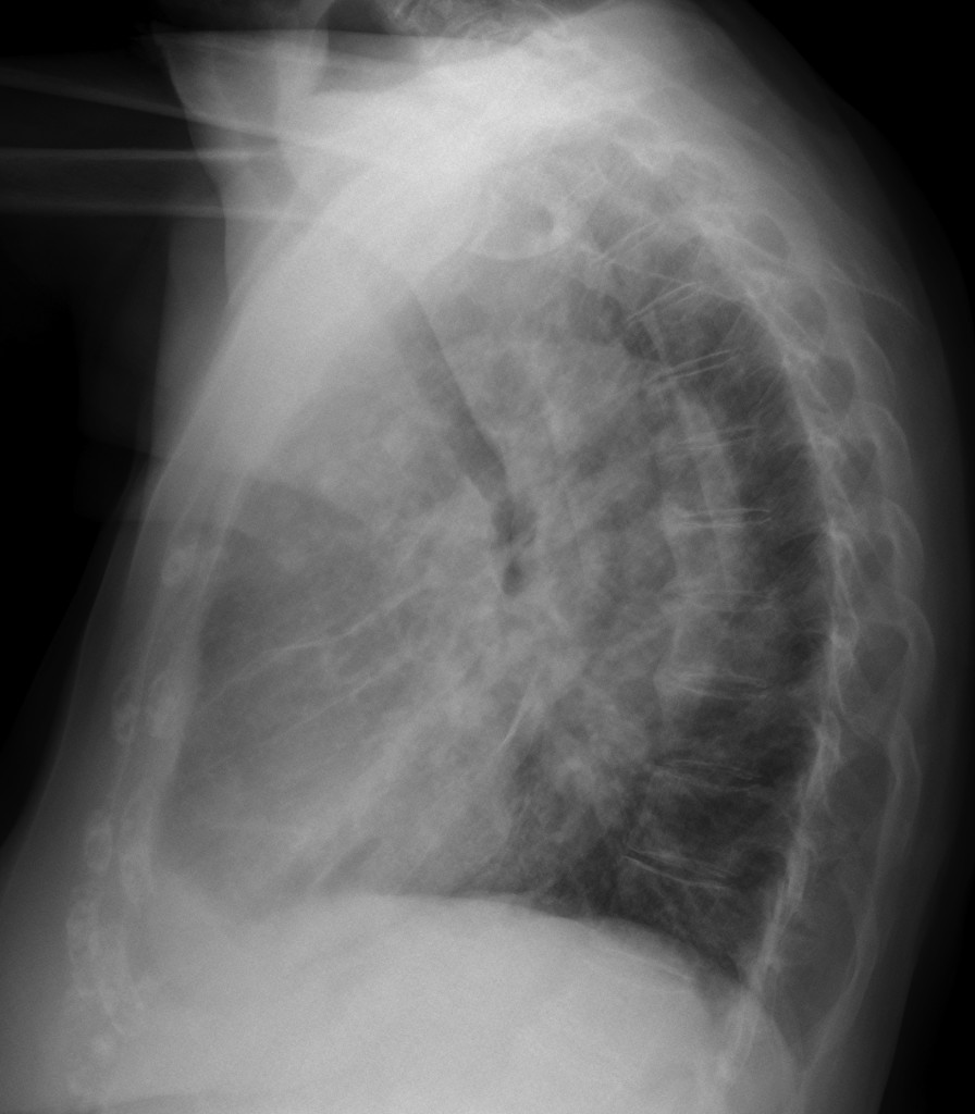
lateral chest, 86-year-old woman
1. Metastases
2. Granulomas
3. Bronchioalveolar carcinoma
4. Lymphoma
Leave your comments below and look out for the answer next week.
Click here for the answer to Case 47
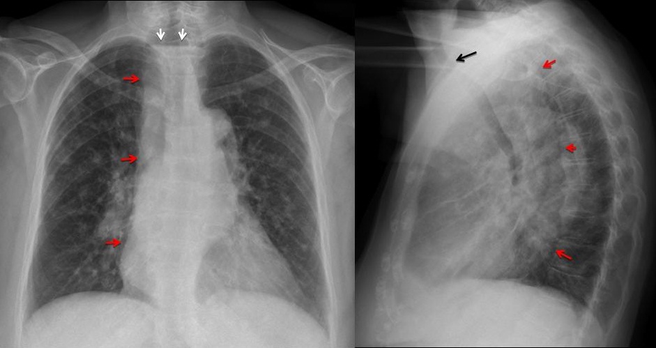
Findings: PA chest radiograph shows an air-fluid level in the upper mediastinum (arrows). There is widening of the right mediastinum (red arrows). On the lateral view there is forward displacement of the trachea (arrow) with occupation of the whole middle mediastinum (red arrows).
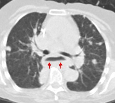
The findings are highly suggestive of a dilated esophagus. In addition, pulmonary nodules can be seen. CT confirms the marked dilatation of the esophagus (arrows) and the pulmonary nodules. This combination suggests two possible etiologies: carcinoma of the distal esophagus with metastases or achalasia with aspiration. In this particular case, the pulmonary lesions did not change for two years, which points to post-aspiration granulomas.
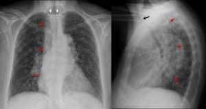
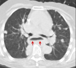
Final diagnosis: achalasia (surgically proven) with pulmonary aspiration, possible atypical TB granulomas (unproven).
Teaching point: this case completes the gamut of air-fluid levels in the chest: mediastinum, pleural space and pulmonary cavities. They are easy to recognize during imaging in the plain film and help to plan the right diagnostic approach.
Muppet wishes very happy holidays to all of you!