Dear friends,
Welcome to case #5.
Case #5 is also from my wife. It is a 27-year-old female with a chronic infiltrate in the right upper lobe. My Wife asked us (ordered, according to Muppet) to help her with the case, because she doesn’t know what it is.
Diagnosis:
1. Tuberculosis
2. Aspiration pneumonia
3. Bronchioalveolar carcinoma
4. Lymphoma
I didn’t make the diagnosis, but the Muppet did! What is the Muppet’s diagnosis?
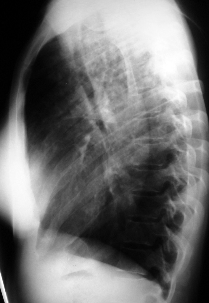
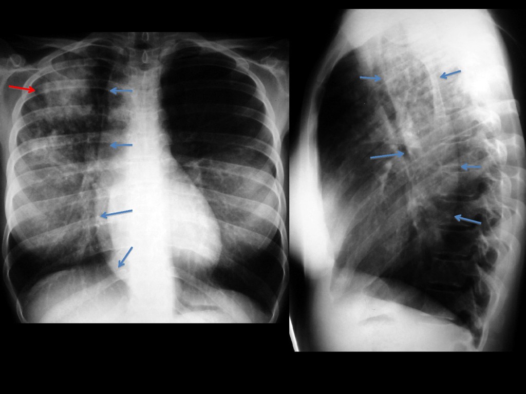
Lateral chest
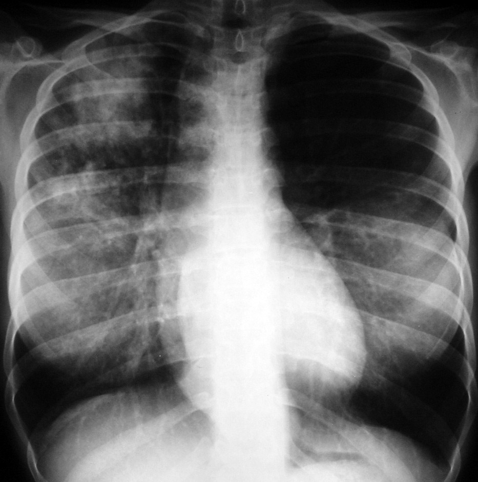
PA chest
N.B. The quality of the films is not excellent, but they give enough information to allow diagnosis. Disregard the increased opacity in both middle lung fields; this is due to overlapping breast prosthesis.
Click here for the answer to case #5
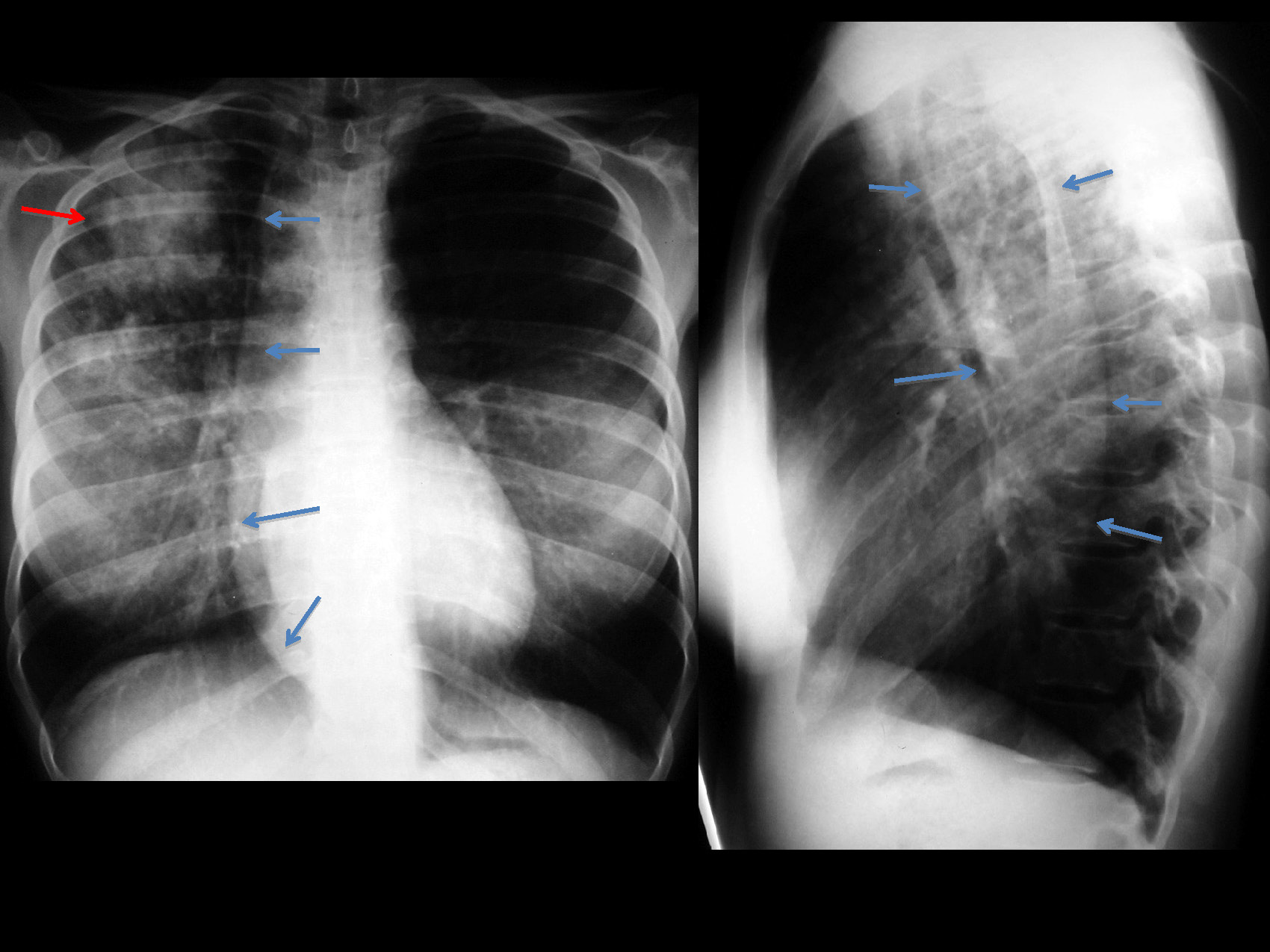
The PA chest radiograph shows an infiltrate in the right upper lobe (
red arrow). There is a right paramediastinal line that extends from top to bottom (
blue arrows). The lateral view shows anterior displacement of the trachea by a tubular structure (
blue arrows), which also extends from top to bottom.
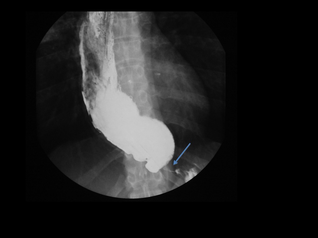
A structure in the middle mediastinum that goes from top to bottom suggests a dilated oesophagus. The anterior displacement of the trachea also supports this possibility. A barium swallow confirms the dilatation of the oesophagus, secondary to narrowing at the oesophagogastric junction (arrow). Best possibilities are carcinoma vs. achalasia. Considering the age of the patient, achalasia with aspiration pneumonia is the most likely diagnosis.
Muppet offers additional information: in achalasia, oesophageal contents may be colonised by nontuberculous Mycobacteria. In a patient with achalasia with chronic pulmonary infiltrates think of infection by nontuberculous Mycobacteria.
Teaching point: Remember that in small children and recumbent patients aspiration pneumonia goes to the right upper lobe.
Muppet insists: avoid satisfaction of search. Don’t be satisfied with initial findings. Keep looking. Not all infiltrates in upper lobes are due to tuberculosis!