Muppet wishes to present the case of a 75-year-old woman with bilateral mastectomies for carcinoma 10 and 7 years previously. Chest radiographs and CT are shown.
1. Pleural metastases
2. Mesothelioma
3. Pleural TB
4. None of the above
Answer case 51
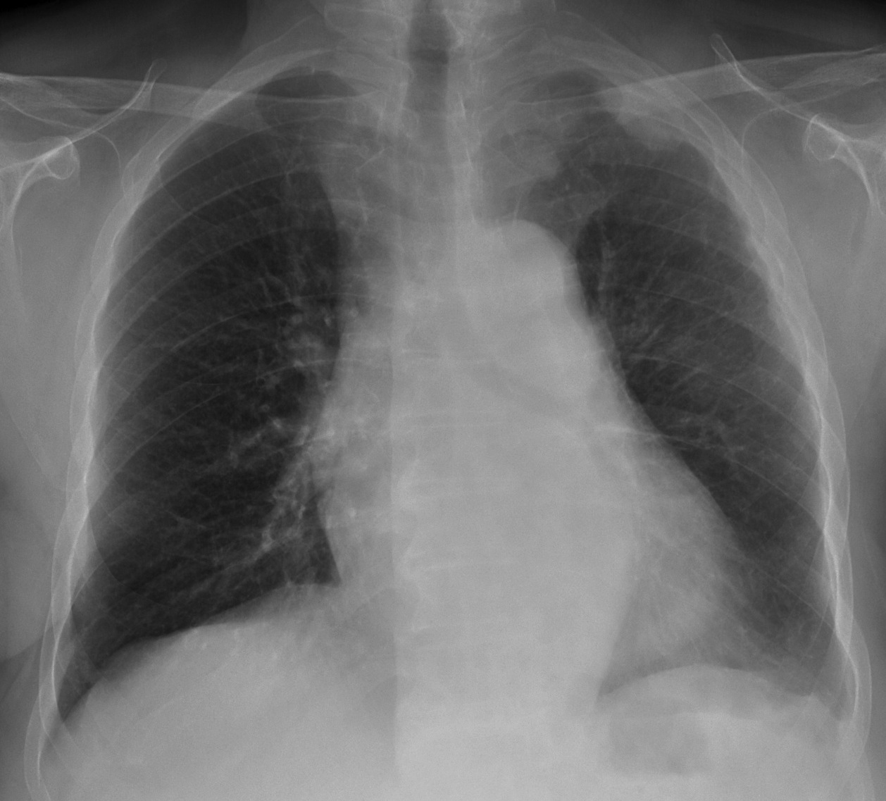
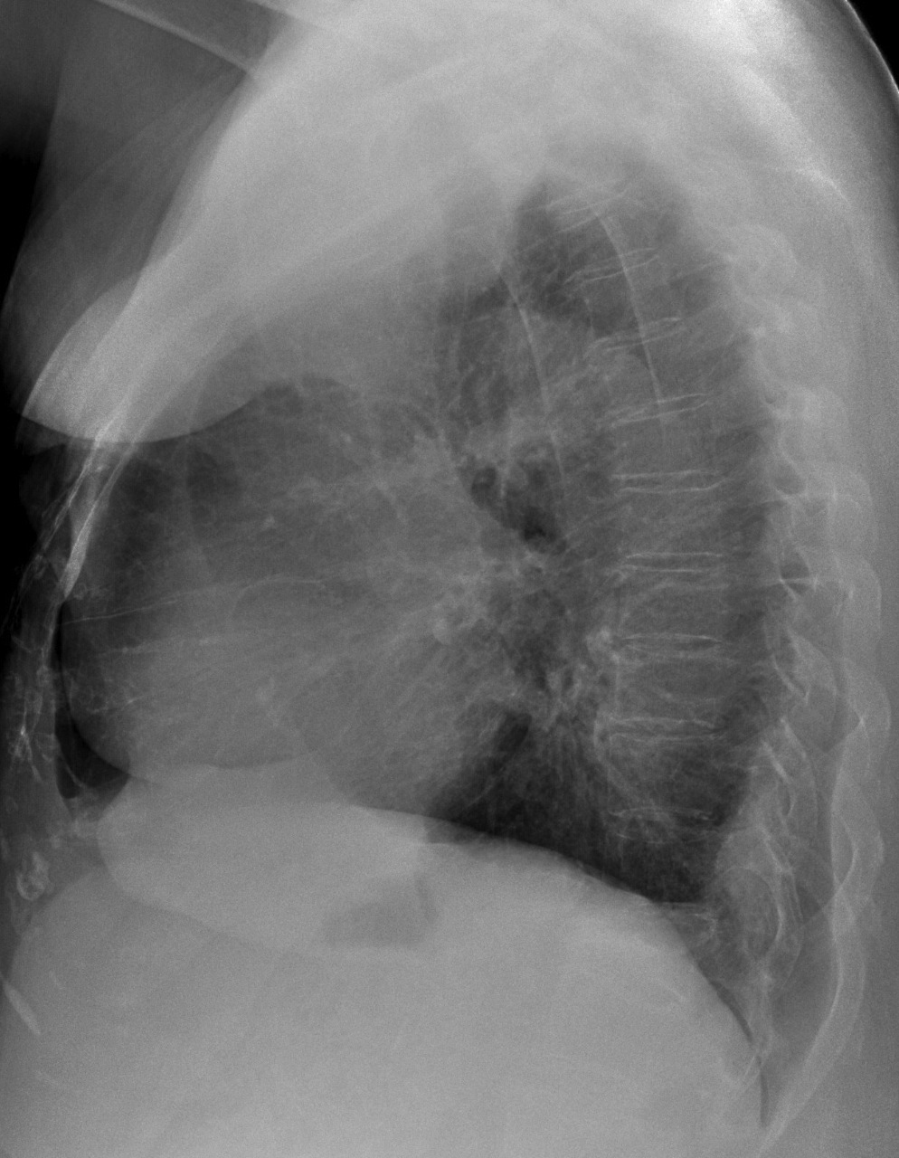
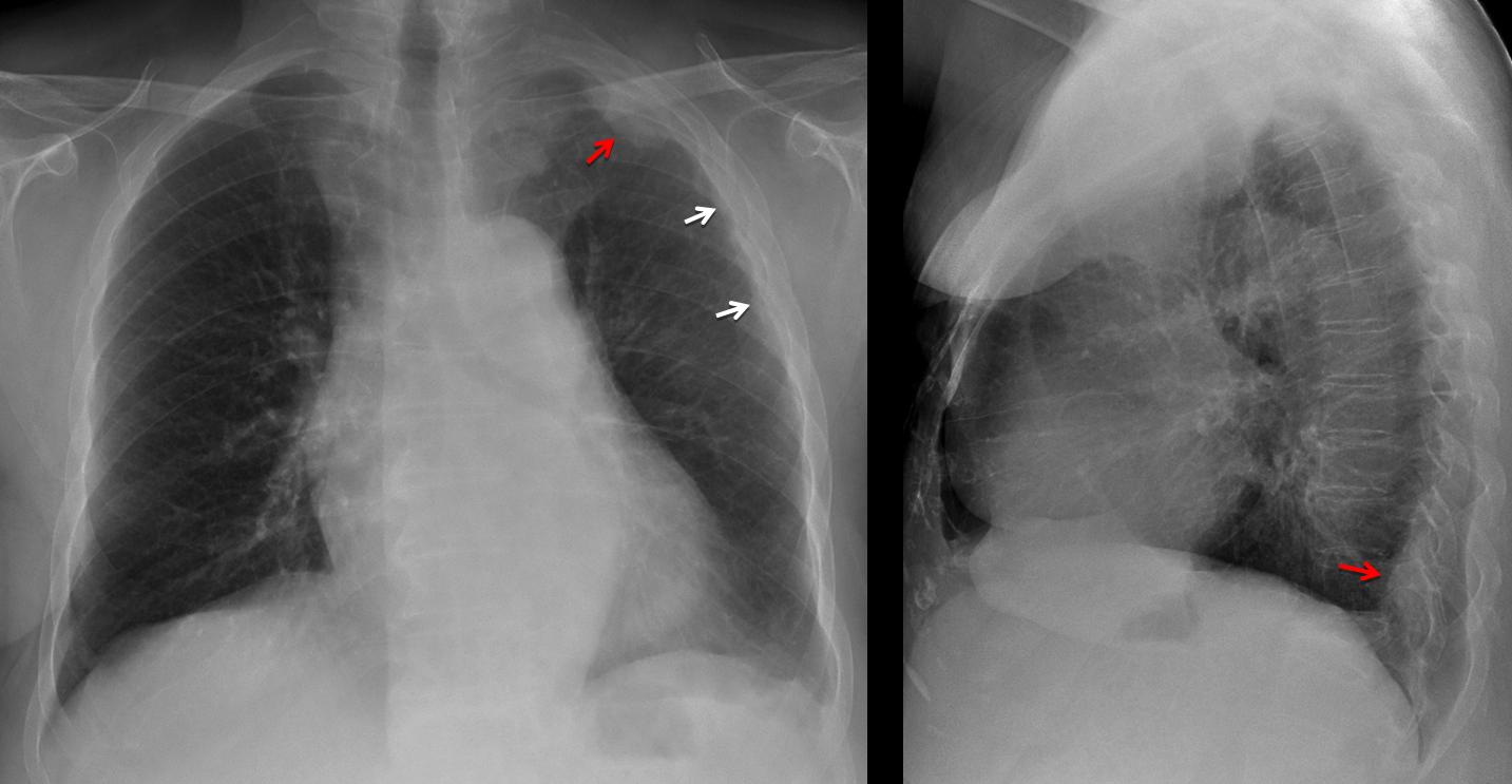
Findings: PA chest shows pleural thickening along the left chest wall (arrows, with blunting of the costophrenic angle and apical thickening. There is also an apical extrapulmonary lesion (red arrow). Lateral film suggests a second lesion (red arrow).
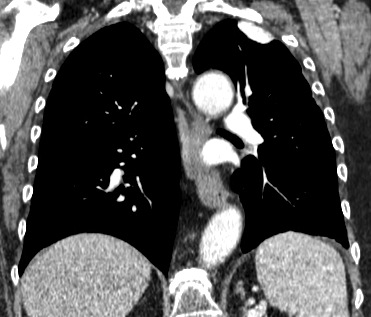
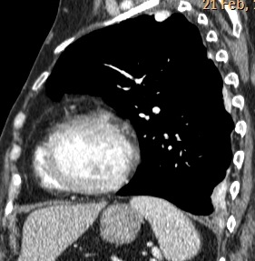
There is collapse of D-12. Sagittal and coronal CT show several high-density pleural-based lesions (arrows).
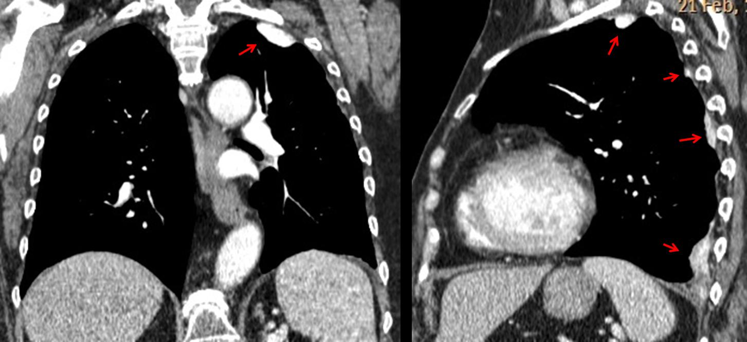
The differential diagnosis includes, among others, asbestos plaques, amyloid, pleural TB and calcified metastases after treatment. In an oncologic patient, another consideration should be talc pleurodesis. This patient had such a procedure in 2008 for recurrent pleural effusion. Appearance of chest two years later was unchanged.
Final diagnosis: talc pleurodesis
Congratulations to Dr. Genchi, who was the first to suggest the diagnosis
Teaching point: any particular finding (in the chest or otherwise) may have a iatrogenic origin. Don’t forget to ask if any procedure has been performed.