Dear Friends,
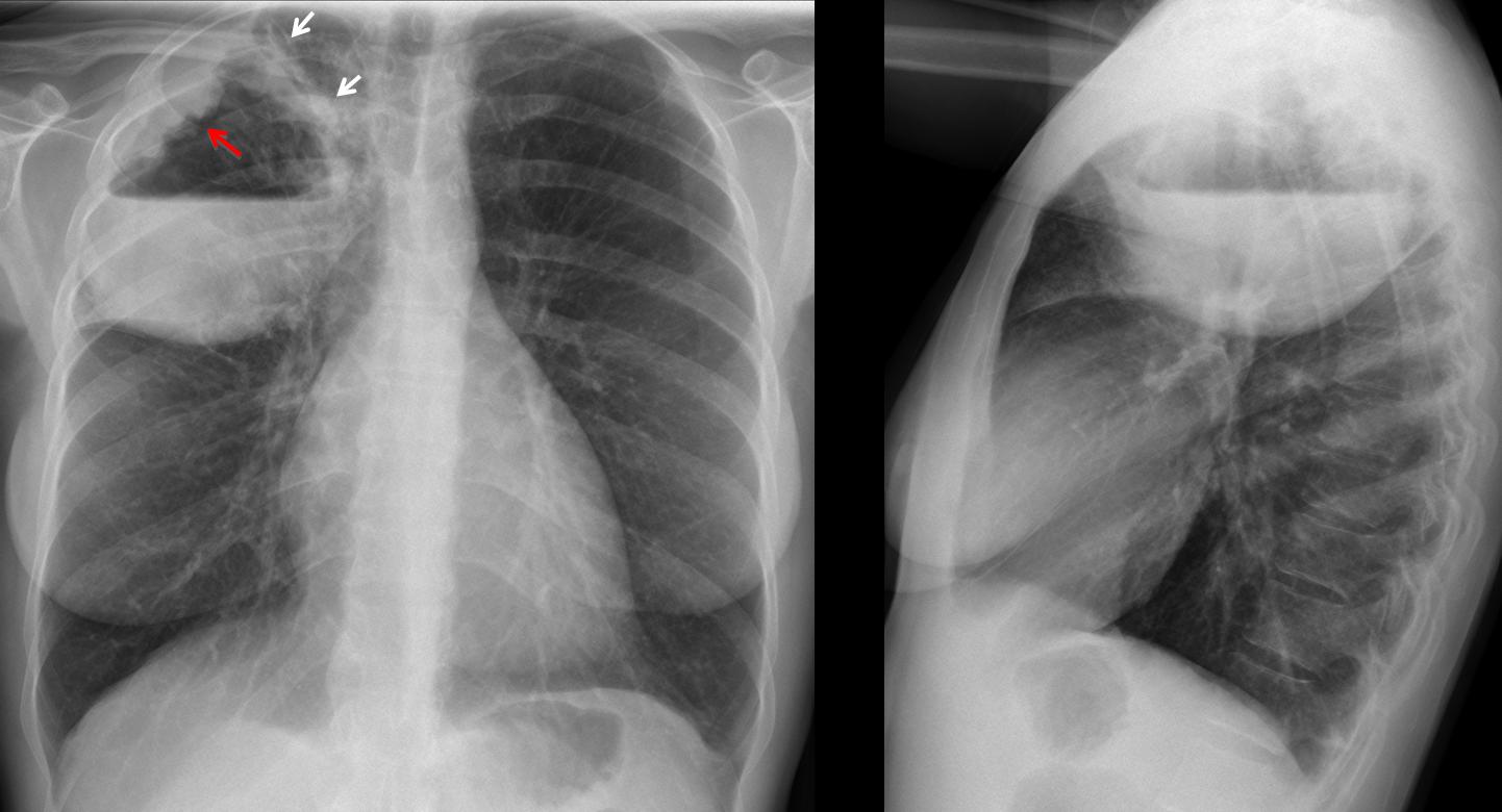
Showing radiographs of a 42-year-old woman with high fever.
Diagnosis:
1. Carcinoma of the lung
2. Pulmonary abscess
3. Loculated empyema
4. None of the above
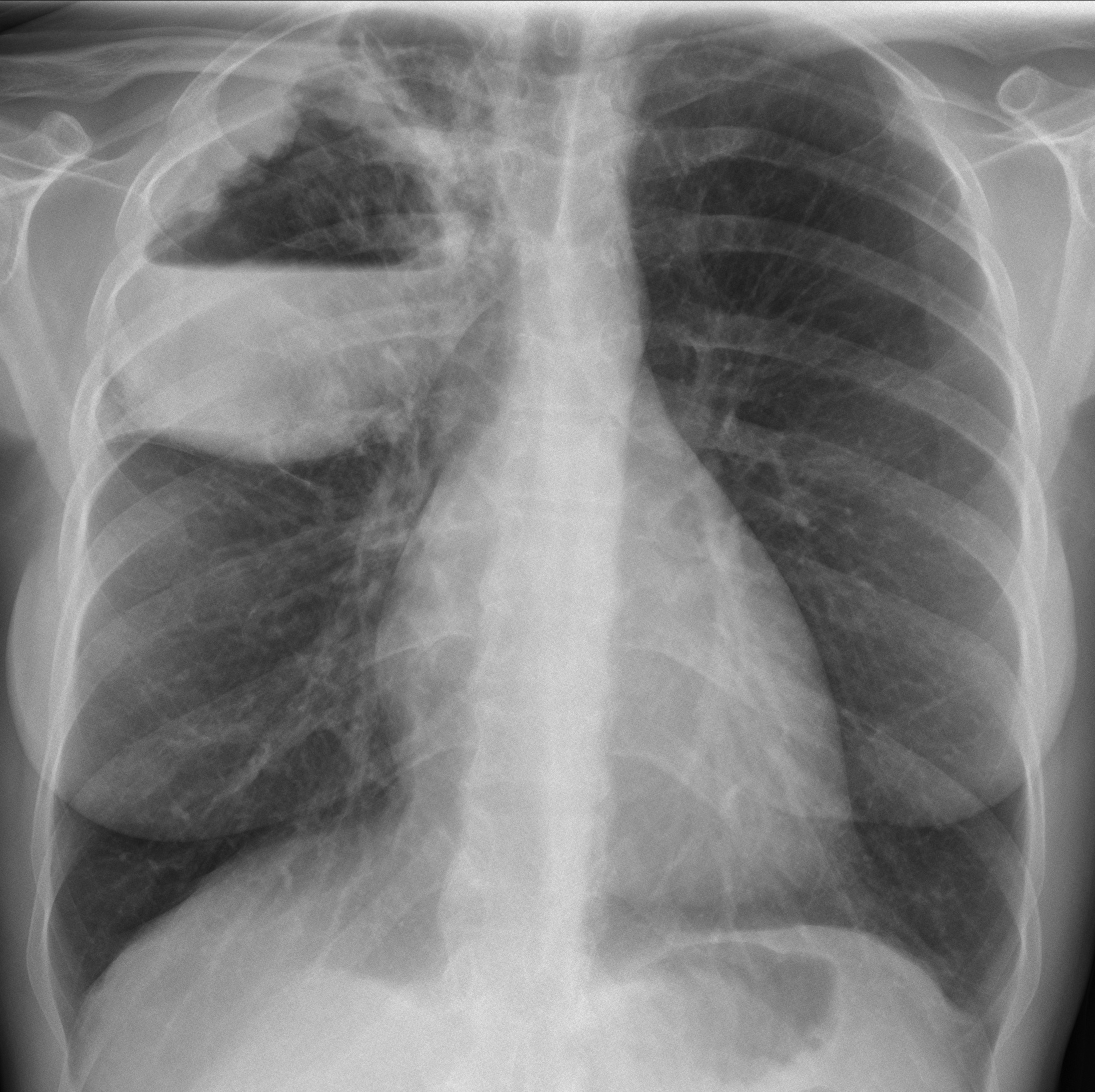
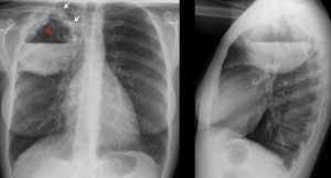
42-year-old woman, PA chest
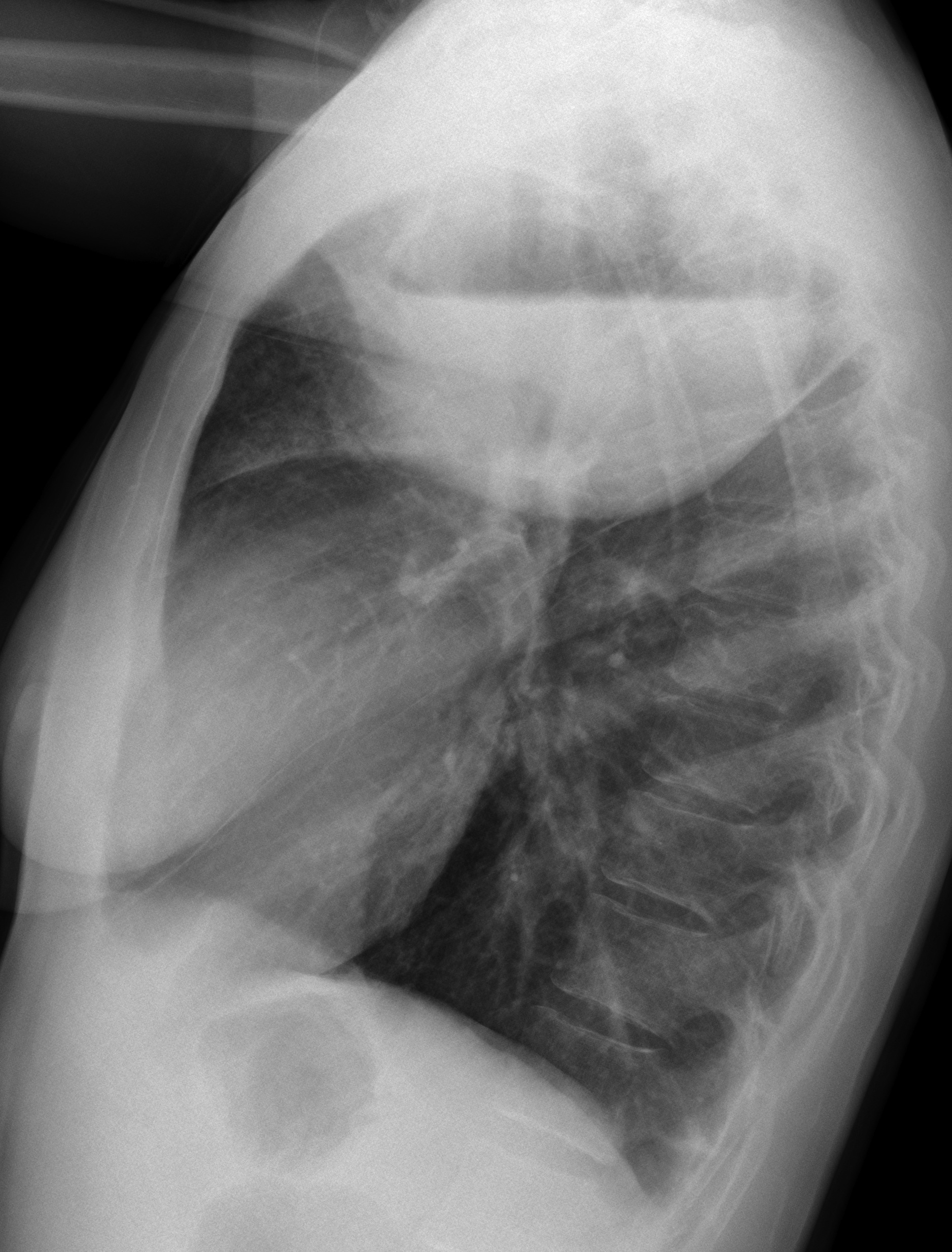
42-year-old woman, lateral chest
Click here for the answer to case #52
Findings: chest radiographs show an RUL lesion with an air-fluid level. The lesion is clearly intrapulmonary because it has an upper wall (arrows) and the air-fluid level is about the same size in both projections. The main finding is an irregular mass protruding from the lateral wall of the cavity (
red arrow). This finding strongly suggests a malignant process.
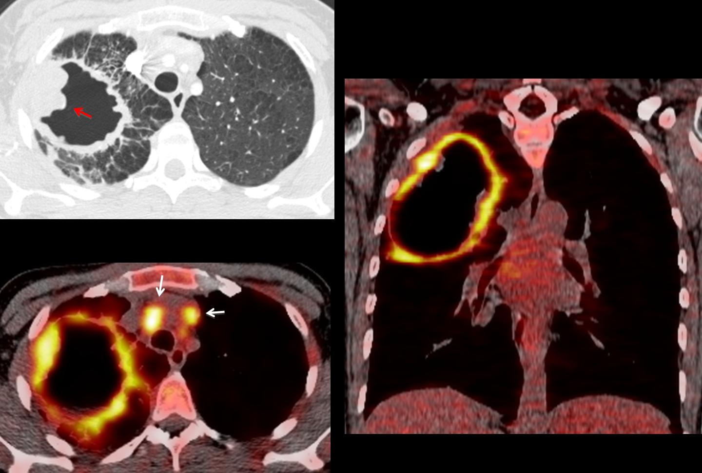
CT confirms the wall mass (red arrow). Additional criteria of malignancy are the numerous Kerley lines around the lesion. PET-CT images show enhancement of the wall and mediastinal lymph nodes (arrows).
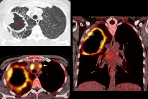
Final diagnosis: infected squamous-cell carcinoma.
Congratulations to Dr. Amutha, who was the first to suggest the right diagnosis.
Teaching point: cavitated inflammatory masses may have irregular walls, but a localised wall mass is highly suspicious of malignancy.