Dear Friends,
My colleague Jordi Andreu, who is almost as mischievous as the Muppet, presents the following case: a 45-year-old woman with pain in the chest.
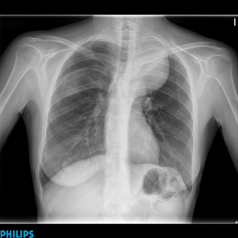
45-year-old woman, PA chest
Diagnosis:
1. Neurogenic tumour
2. Thymoma
3. Fibrous tumour of pleura
4. None of the above
Click here for the answer to case #54
This case shows interesting semiologic findings. There is an obvious left mediastinal mass, which extends to the lung apex, placing the mass in the posterior mediastinum. It cannot be a thymoma because the upper mediastinum ends at the level of the clavicles. A goiter can be excluded because the trachea is not displaced. In addition, there is remodelling of the right fifth rib (arrows) and ribbon-like appearance of the right and left third ribs (arrows). All these findings point to neurogenic tumours in a patient with neurofibromatosis.
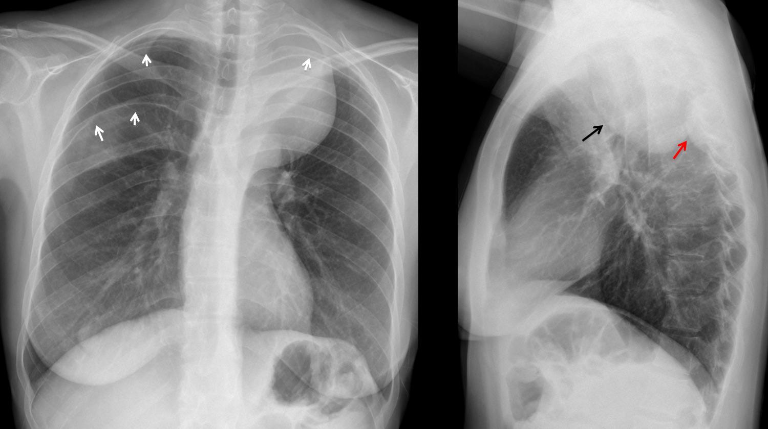
The lateral view shows the large mass (arrow) and a smaller one (red arrow), which is responsible for the remodelling of the right fifth rib. CT confirms the presence of both masses (arrows).
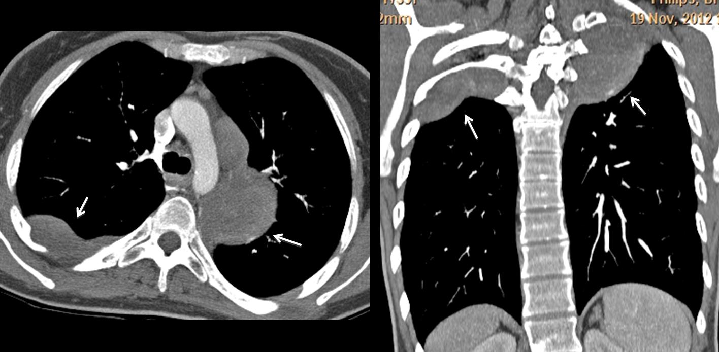
Remodelling of the right fifth rib is better demonstrated in the 3D reconstruction (arrows). PET-CT shows high uptake of the left mass.
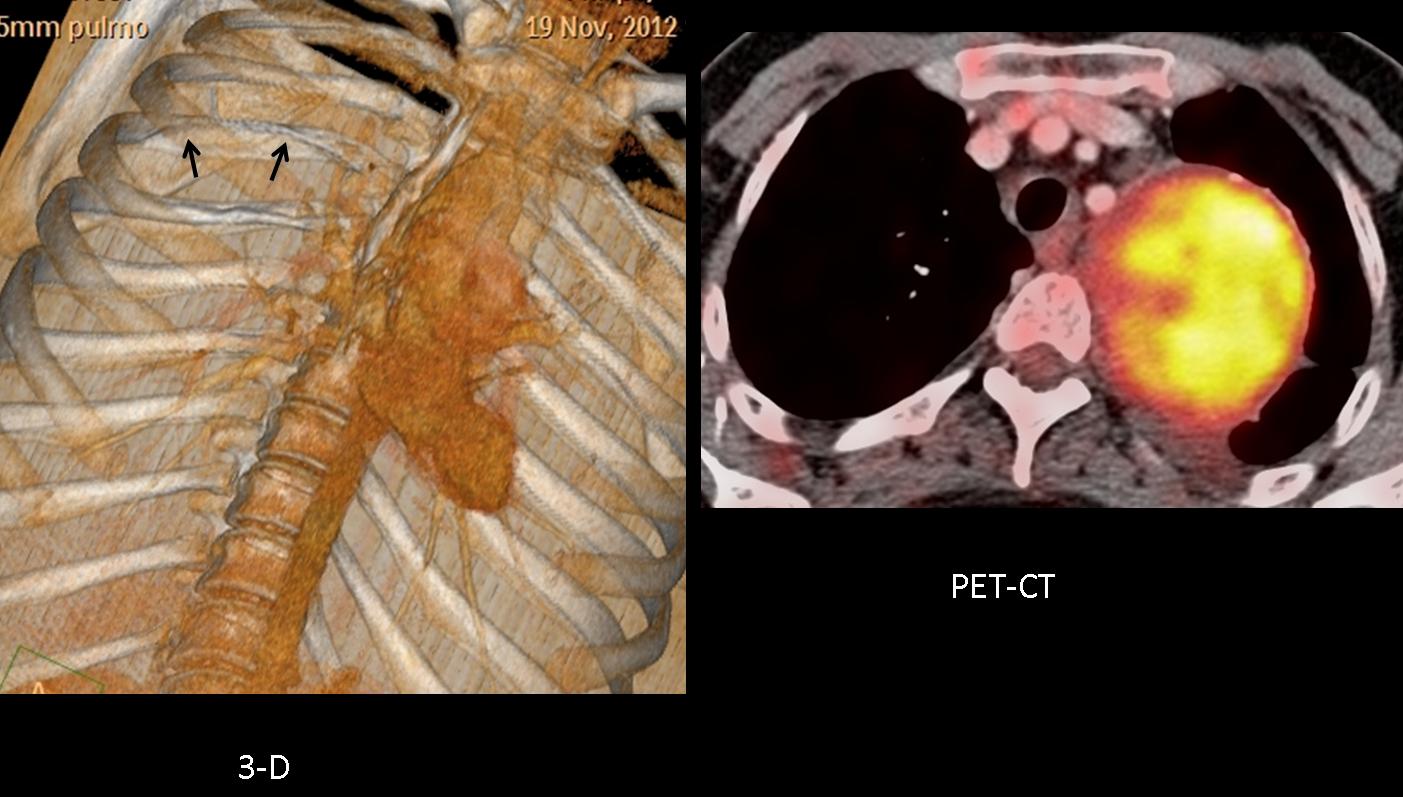
Final diagnosis: neurofibromatosis type I, with malignant degeneration of the big left chest tumour.
Teaching point: posterior mediastinal masses accompanied by rib notching and ribbon-like ribs are highly suspicious of neurofibromatosis.


