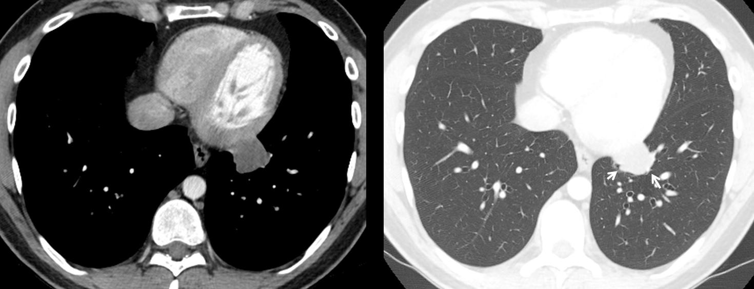
Showing images of a 30-year-old male, asymptomatic. What would be your diagnosis?
1. Pericardial cyst
2. Lung tumour
3. TB lymph nodes
4. None of the above
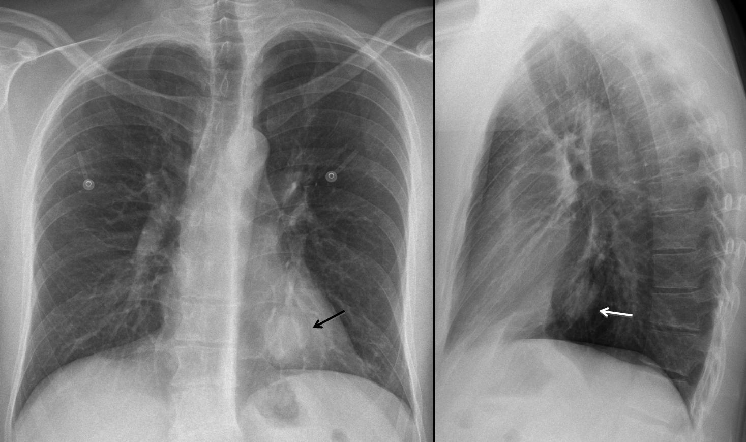
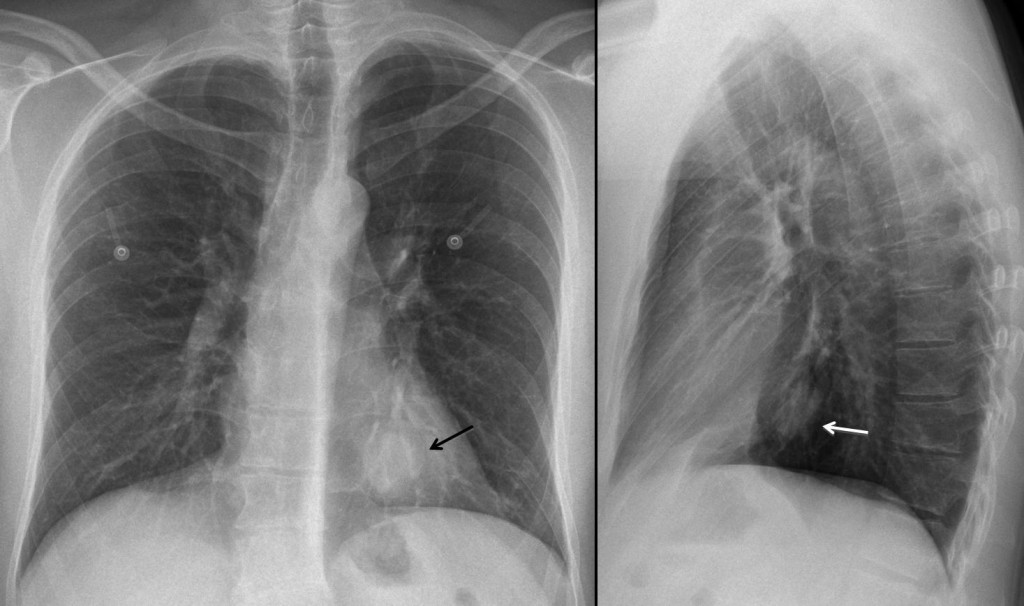
Findings: PA and lateral chest show a rounded lesion behind the cardiac silhouette (arrows).
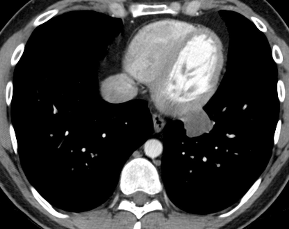
CT shows a slightly irregular hypodense mass, which does not enhance with IV contrast.
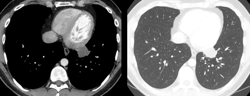
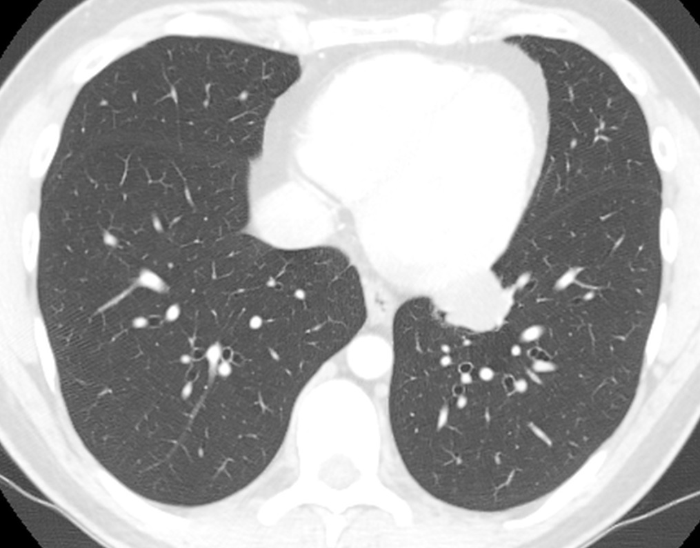
In the pulmonary window the mass is poorly defined, with irregular borders (arrows). This combination of signs speaks against a mediastinal mass and makes the lesion intrapulmonary, excluding pericardial cyst and mediastinal lymph nodes.
Final diagnosis: mucinous adenocarcinoma of the lung (explaining the hypodensity of the mass).
Congratulations to Dr. Prasanna Kulkarni who made an accurate description of the findings and arrived at the correct conclusion.
Teaching point: recognising the signs that differentiate intra from extrapulmonary lesions will lead you to the right diagnosis, as Dr. P. K. did.