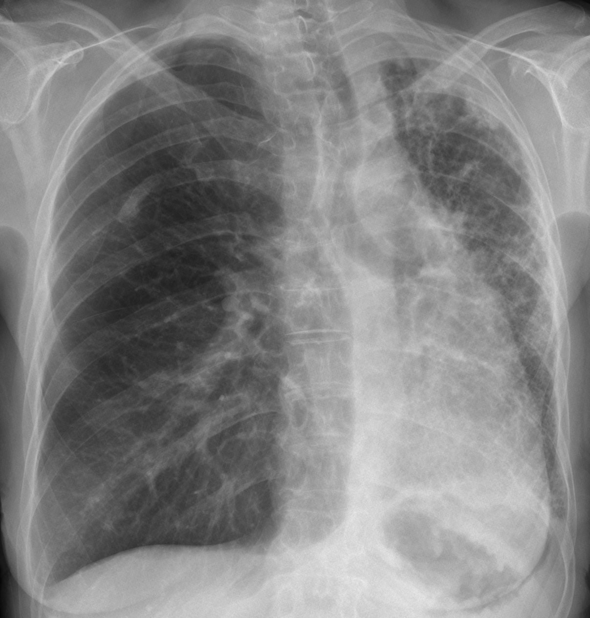
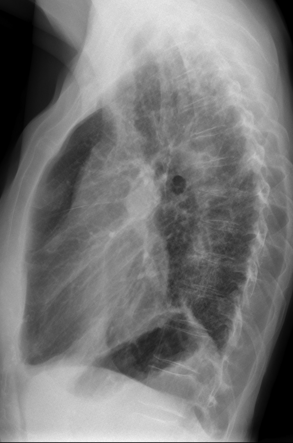
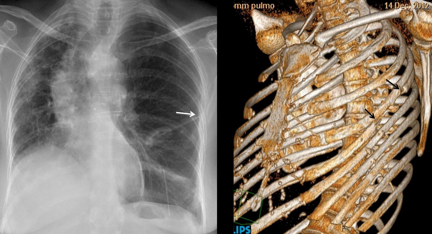
Muppet cannot find difficult cases and, despite himself, is forced to show easy ones. Today we’re showing radiographs of a 48-year-old woman who has had moderate dyspnea for a while.
1. Swyer-James syndrome
2. Tumour left main bronchus
3. Old TB left lung
4. None of the above
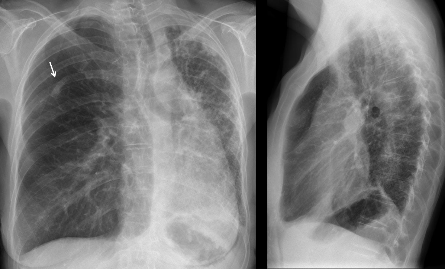
Findings: PA and lateral radiographs show a small left hemithorax with diffuse lung disease. Right lung is expanded and mediastinal structures are displaced towards the left. Obviously, the diseased lung is the left one. This raises the question of why the right lung is spared. And the answer is very simple: the right lung is OK because it is transplanted. A clue to the diagnosis is the surgical fracture of the right 6th rib (arrow).
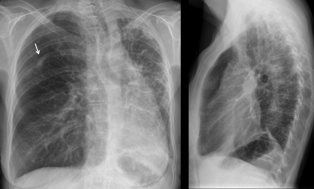
Fig. 1
I wanted to show the typical appearance of a condition that it is not uncommon. About 4,000 lung transplants are performed each year; 1,677 of them in the European Union. Knowing this appearance (they all look the same, see Fig. 2) will prevent errors, especially if you are taking an examination.
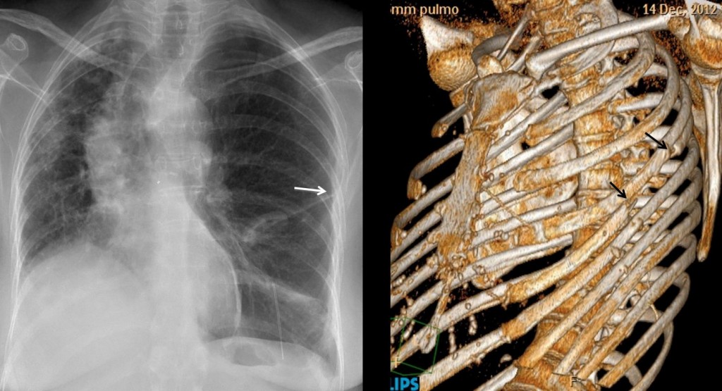
Fig. 2. Left lung transplant. Note post-surgical changes in the left 5th rib (arrows)
Final diagnosis: right lung transplant
Teaching point: When seeing a unilateral hyperlucent lung, consider a previous transplant.
In any patient, isolated fractures of 5th or 6th ribs should raise the question of previous surgery.