Caceres’s Corner Case 57 (Update: Solution)
Dear Friends,
Dr. Manel Martínez, a long-time friend of the Muppet, has provided the following case: an asymptomatic 67-year-old man in whom a mass was discovered in the chest radiographs.
Diagnosis:
1. Aortic aneurysm
2. Hydatic cyst of lung
3. Left pulmonary artery aneurysm
4. None of the above
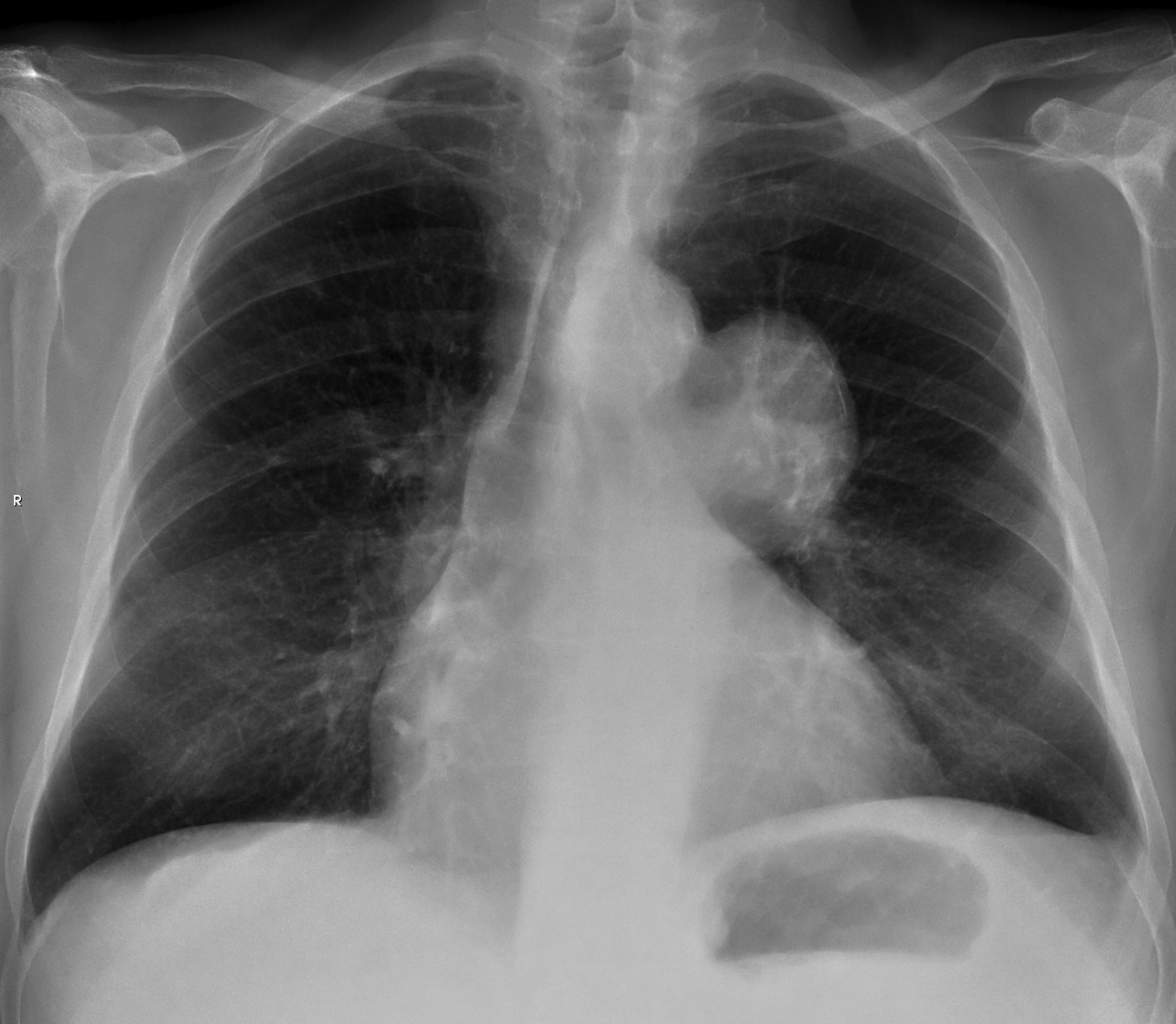
PA chest, asymptomatic 67-year-old man
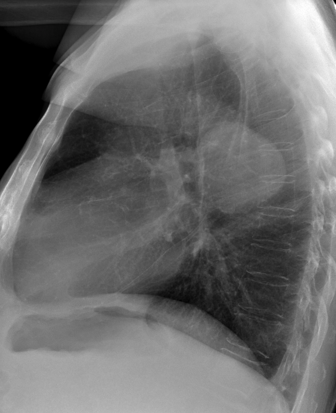
lateral chest, asymptomatic 67-year-old man
Click here for the answer to case #56
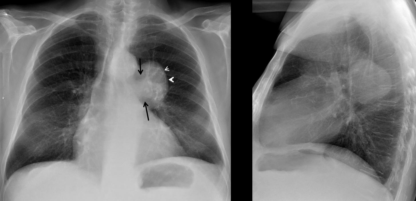
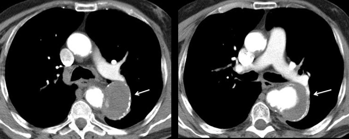
Final diagnosis: saccular aneurysm of descending aorta.
Teaching point: when seeing lineal peripheral calcification, think of cyst vs. vascular (Dr. Pepe dixit).


