Caceres’s Corner Case 57 (Update: Solution)
Dear Friends,
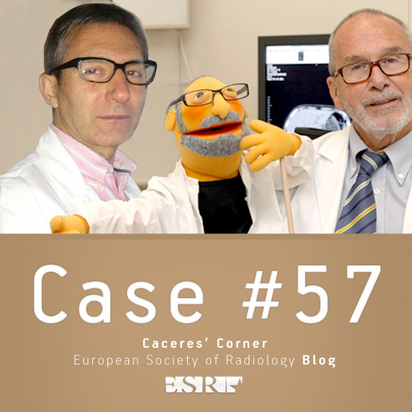
Dr. Manel Martínez, a long-time friend of the Muppet, has provided the following case: an asymptomatic 67-year-old man in whom a mass was discovered in the chest radiographs.
Diagnosis:
1. Aortic aneurysm
2. Hydatic cyst of lung
3. Left pulmonary artery aneurysm
4. None of the above
PA chest, asymptomatic 67-year-old man
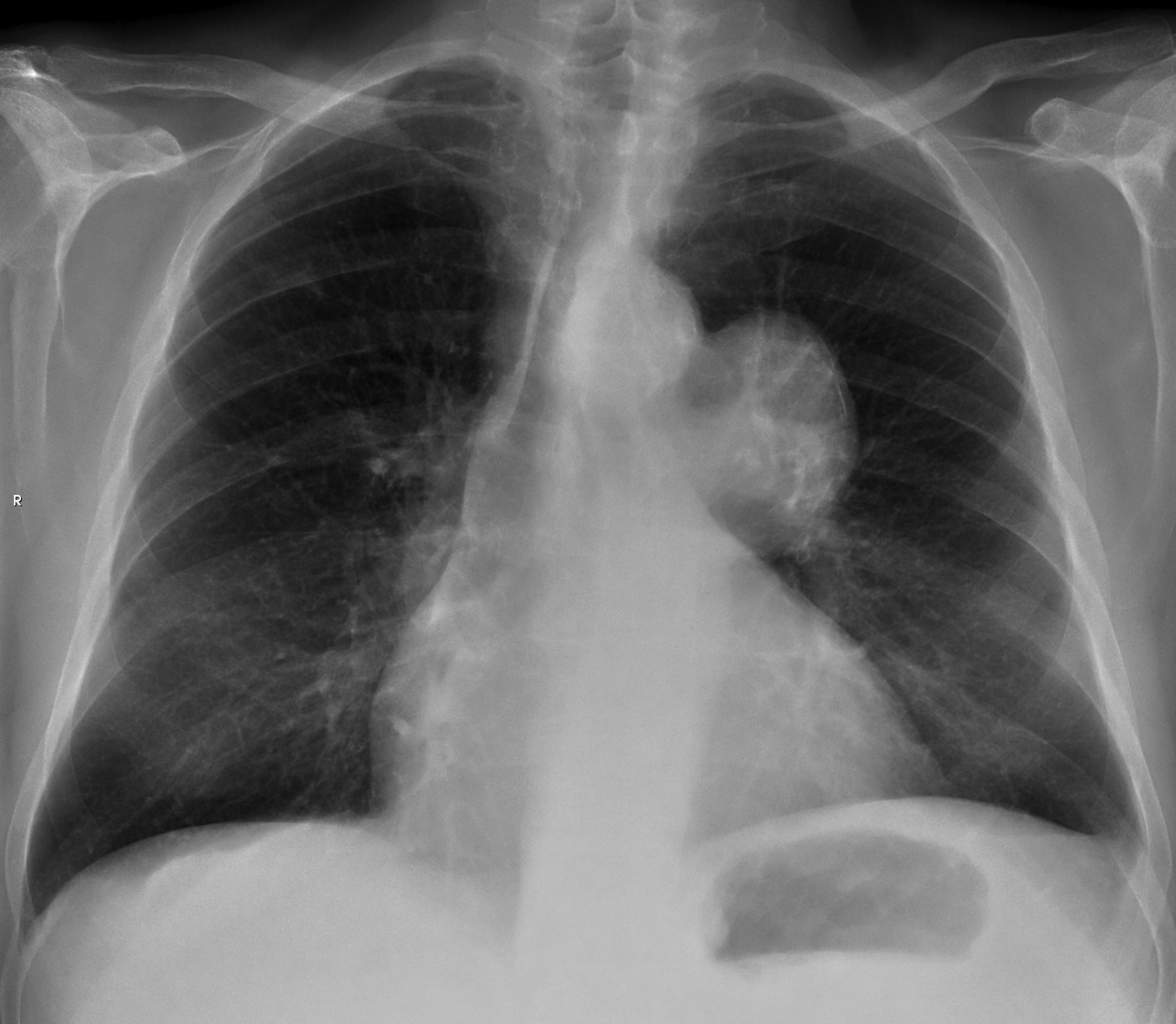
lateral chest, asymptomatic 67-year-old man
Click here for the answer to case #56
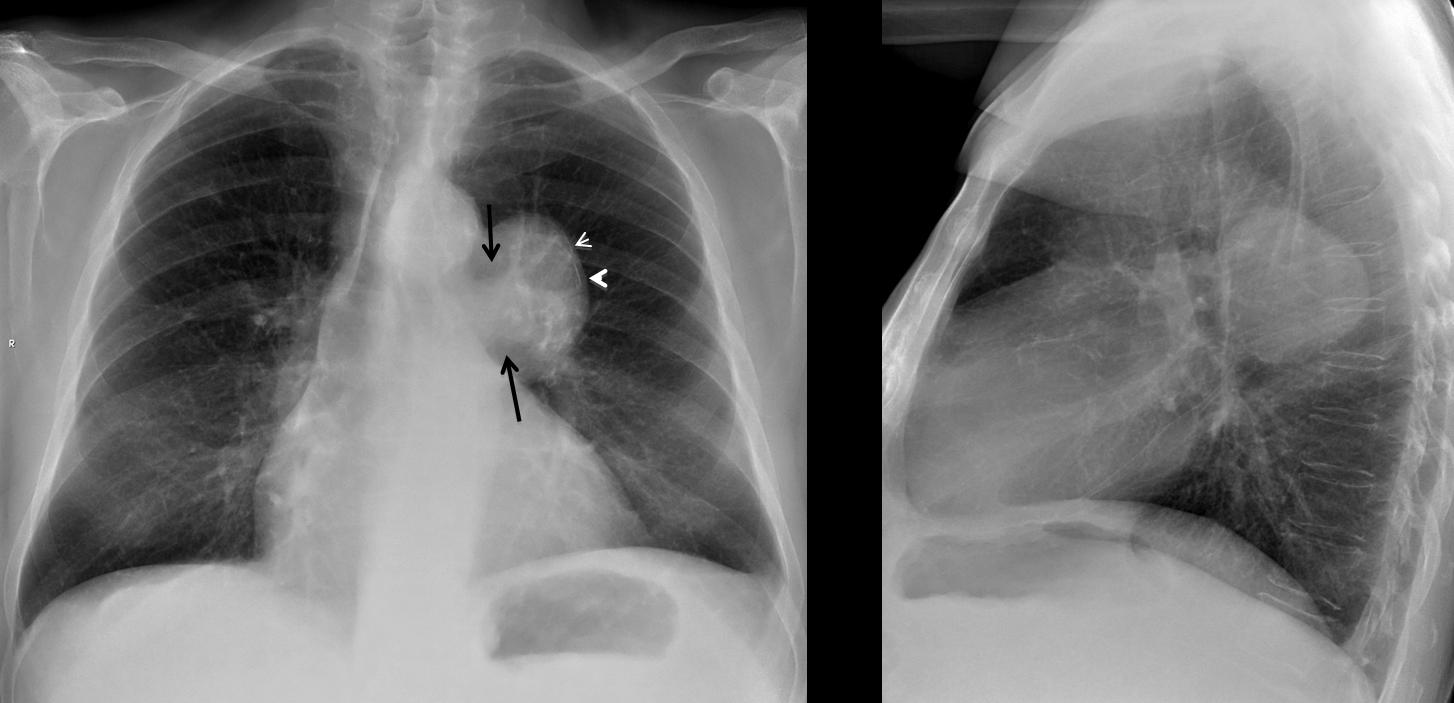
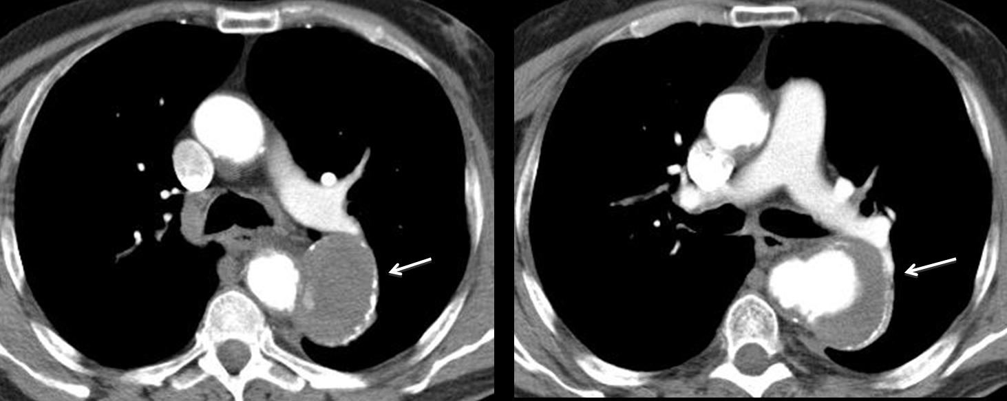
Final diagnosis: saccular aneurysm of descending aorta.
Teaching point: when seeing lineal peripheral calcification, think of cyst vs. vascular (Dr. Pepe dixit).


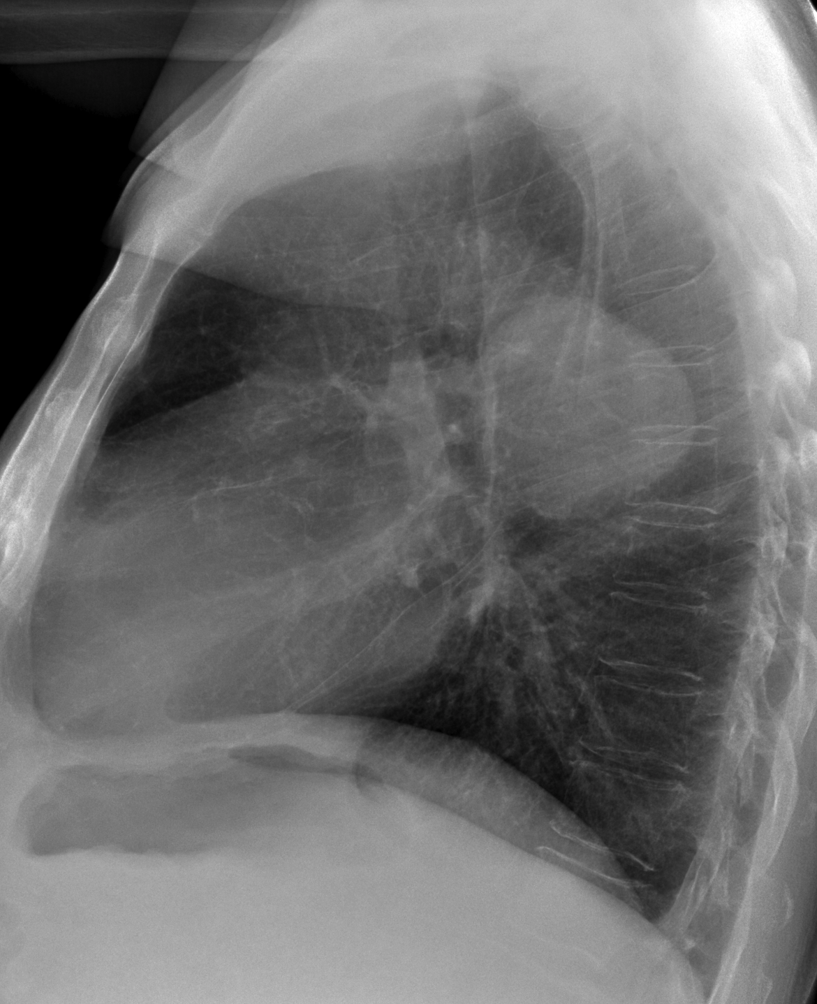
thoracic descending aortic saccular aneurysm
Saccular image that identifies edge mediastinal deforms under the second left aortic arch. Finding suggestive of left pulmonary artery aneurysm
“hilar convergence” and “hilum overlay” signs buddy ?
It is an aspect vascular mediastinal mass that appears to originate in the left pulmonary artery.
True, it is very rare but I consider it an aneurysm of the left pulmonary artery.
Sorry for my bad English.
Rectified, thanks for your comment. It certainly seems to have no hilar origin. Anyway I have my doubts that originates in the aorta.
Still think …………………….
CXR , frontal and lateral views : a large well defined mass is seen projected overlying left hilum , left pulmonary artery is seen clearly , so this mass is either anterior or posterior to the left hilum , lateral view revealed posterior location to the hilum .
aortic contour is intact , not dilated in 67 years old male but localized saccular aneurysm is a possibility .
another possibility to be hydatid cyst especially , presence of thin calcific rim along left lateral border , another possibility in asymptomatic case is bronchogenic cyst . another possibility is posterior mediastinal mass as neurogenic tumor .
so CT scan is advised .
Nice discussion, but I disagree about the rim calcification in hydatid cyst of the lung.
hydatid cysts of lung typically never calcify
ok
hydatid cyst in the lung has no calcified rim .
ok
hydatid cyst in the lung has no calcified rim .
Hello,
If we apply” hilar convergence sign”, we are not supposed to see left pulmonary artery behind the mass if the mass has a vascular origin, isn’t it?
in this case, we can clearly see LPA and its branches behind the mediastinal opacity ; besides the thin calcified rim can be highly suggestive of mediastinal hydatic cyst , that can take place in any mediastinal compartment .
so it should be the most likely diagnosis according to me…
Señor Pepe dijo hace unas semanas – “A very important finding is the discovery of peripheral linear calcification. This type of calcification points to two possibilities:
a cystic lesion or a vascular structure.”
Hence, I go with bronchogenic cyst, since aorta and left pulmonary artery are not obliterated, and hydatid cysts don’t tend to calcify when in the lung.
Dr. Pepe thanks you for remembering his advice. Discovering he linear calcification gives you a 50/50 chance.
Why do you say that the aorta is not obliterated? Do you see the line of the descending aorta?
Saccular descending aortic aneurysm
broncogenic cyst
L’opacità è nel mediastino medio.Essa ha limiti netti, con sottile linea calcifica esterna e “spot” calcifici interni.L’aorta e l’arteria polomonare sx sono chiaramente visibili ed indenni; una cisti idatidea non ha una sede perilare e non contiene calcificazioni.L’ipotesi diagnosticaKISS è una cisti broncogena mediastinica, adesa al bronco principale.Il paziente è asintomatico.
It could very well be a duplication cyst, but with the KISS hypothesis I would say an aortic aneurysm should be more common.
habemus “Messi””: complimenti per il caso e la “remuntada”!Ora vi “apetta” la Iuventus !