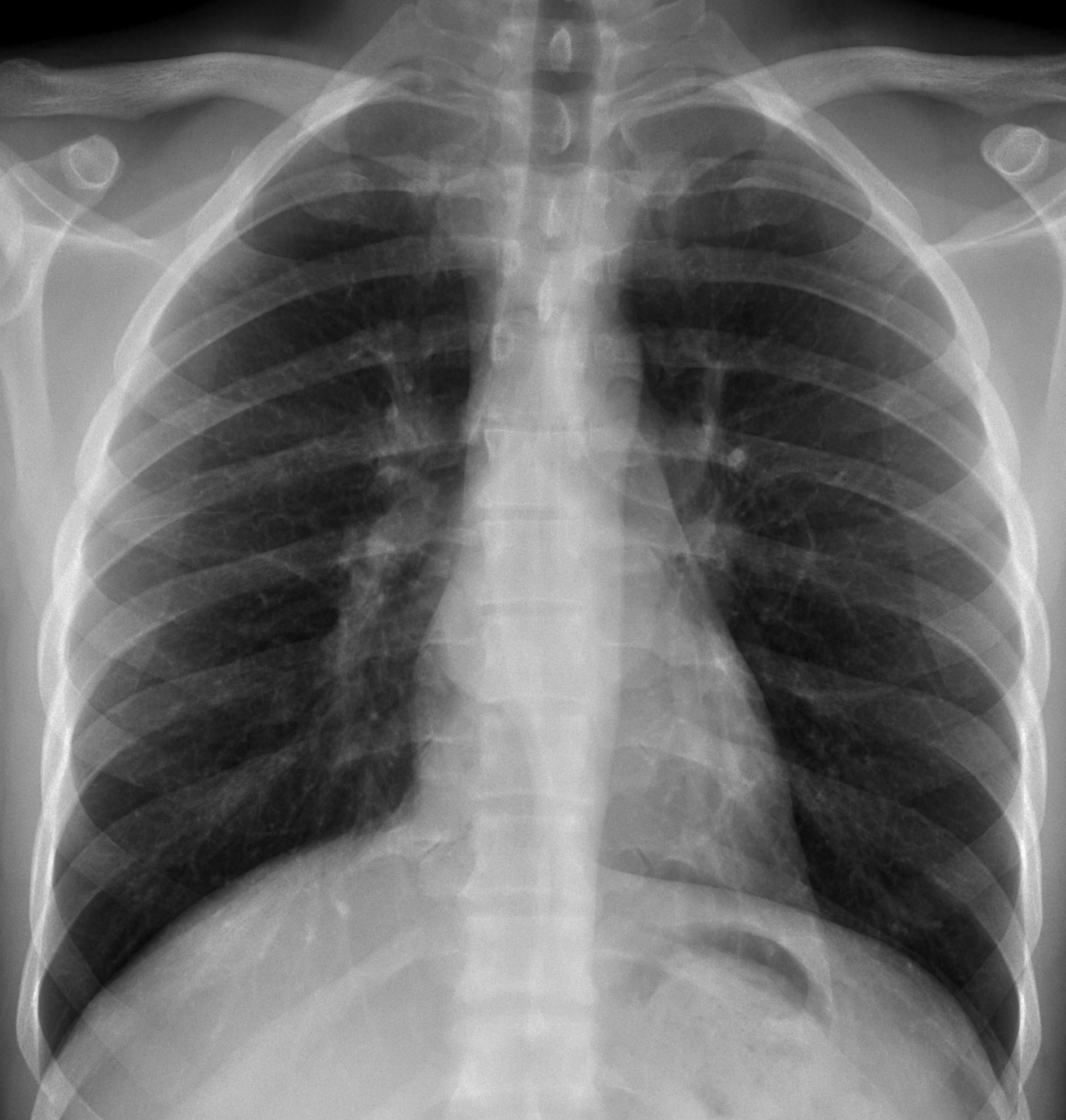
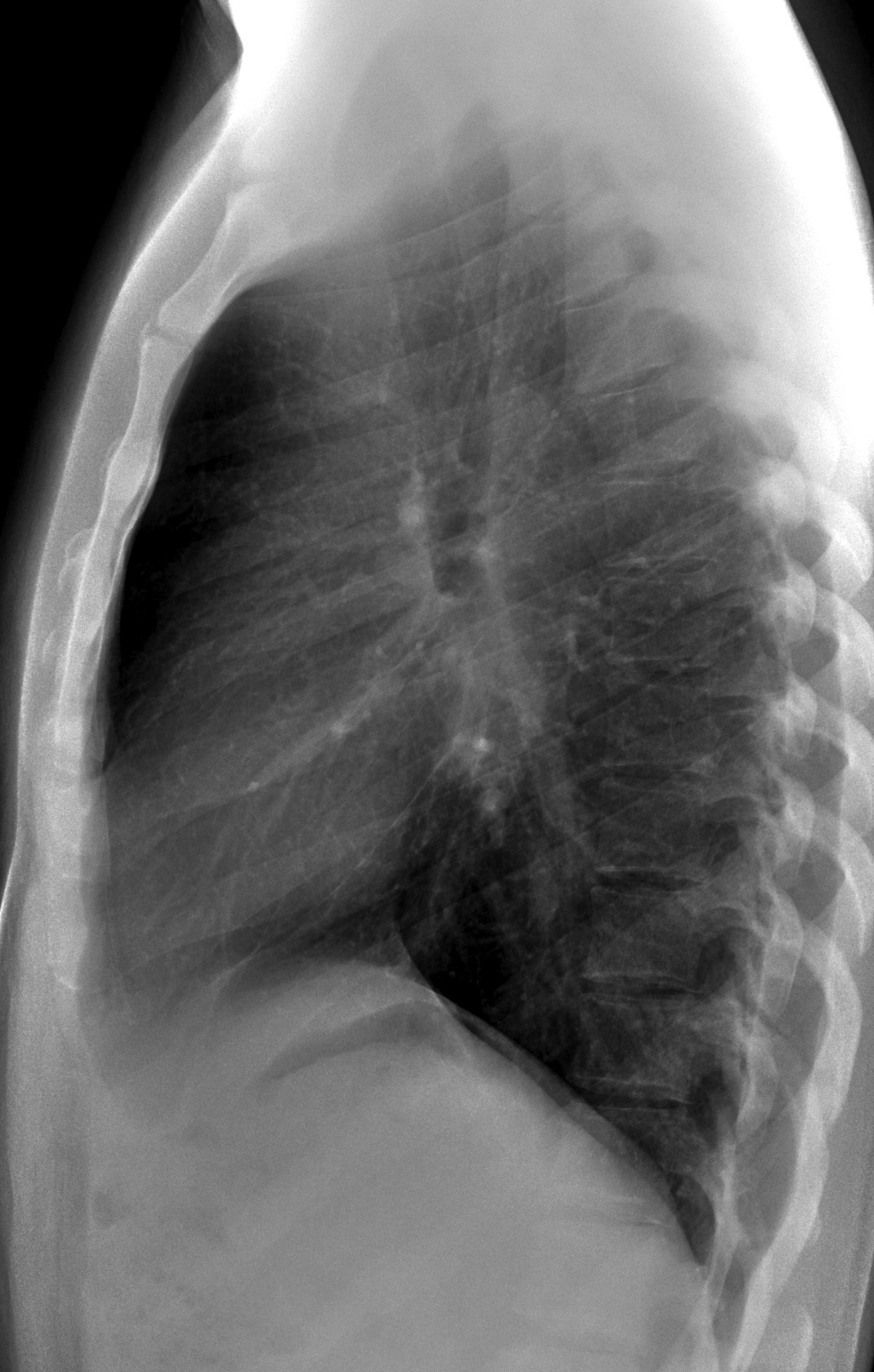
Today, we’re showing you radiographs of a 29-year-old non-European male with moderate dysphagia. Questions:
1. Where is the lesion?
2. What would be your diagnosis?
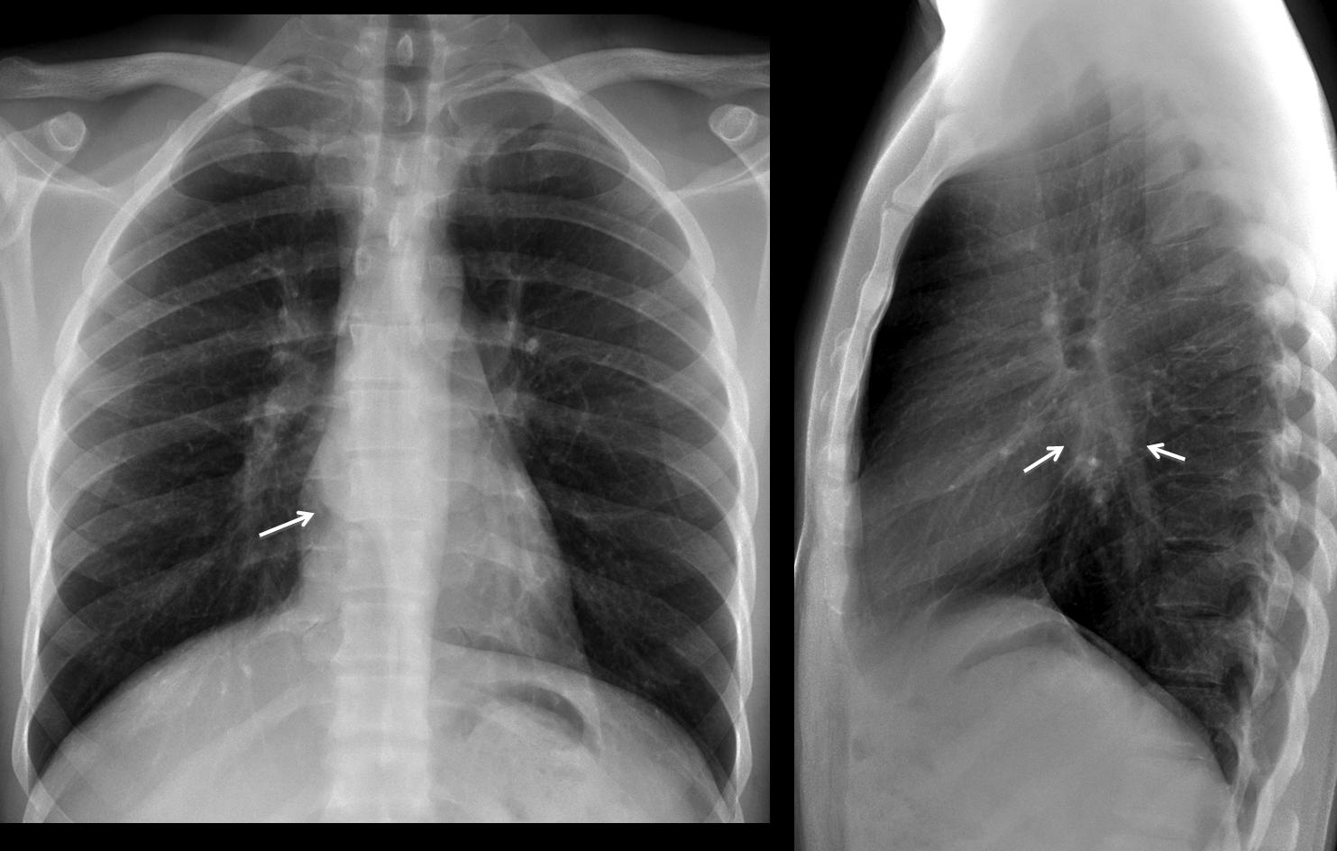
Findings: chest radiographs show bulging of the azygo-oesophageal line in the PA view (arrow) and occupation of the subcarinal space in the lateral view (arrows).
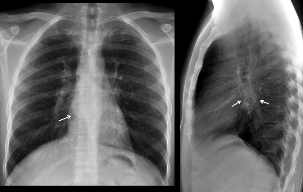
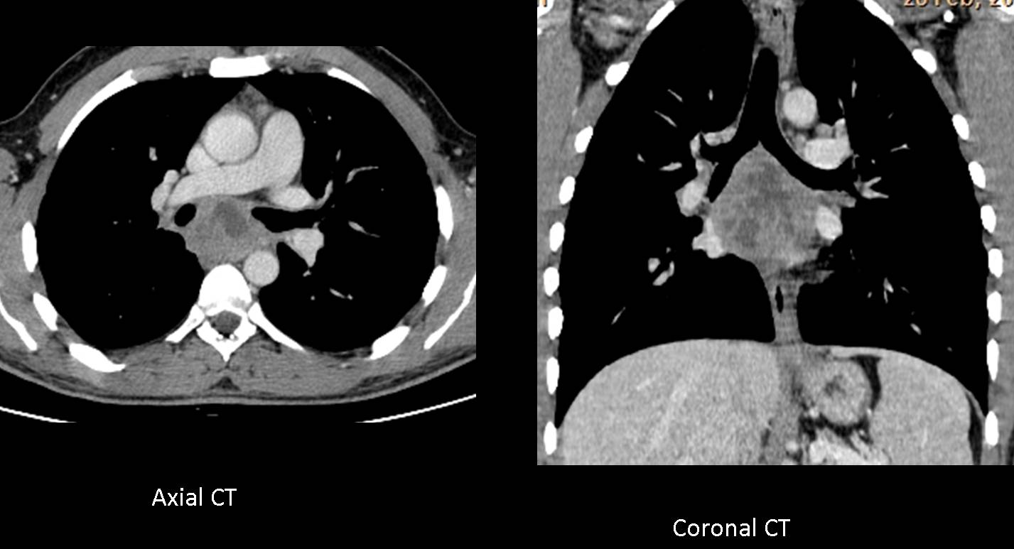
These findings point to a mediastinal process in the middle portion of the middle mediastinum. The most common pathologies in this location are duplication cysts or those related to enlarged lymph nodes. Axial and coronal enhanced CT show an irregular necrotic mass in the subcarinal area, compatible with necrotic lymph nodes.
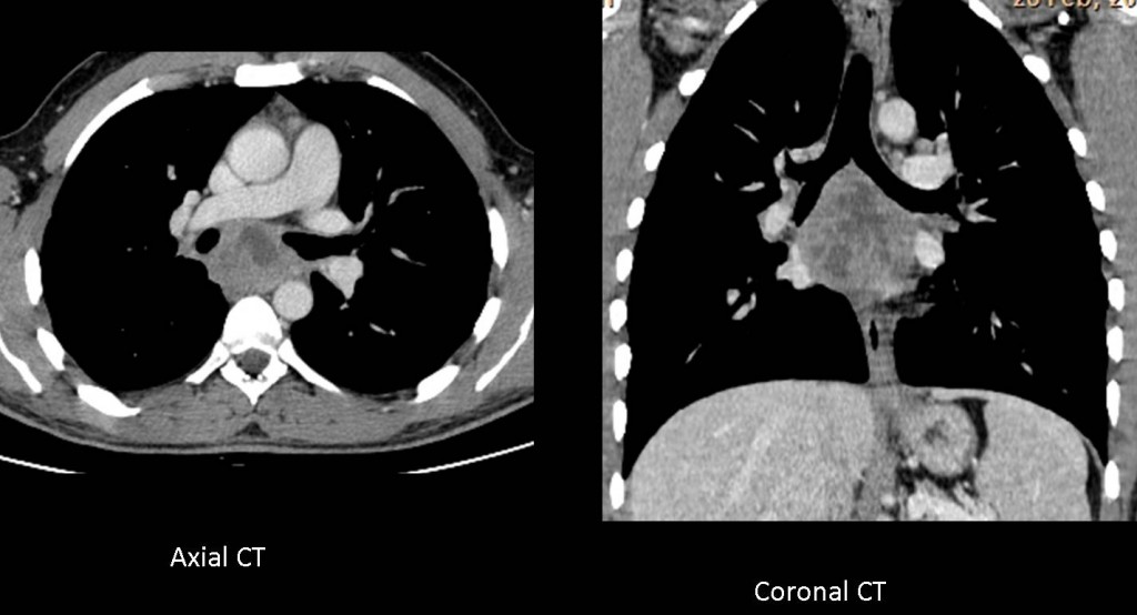
Considering that the patient is non-European, tuberculosis is a strong possibility. PPD was negative, but BK was found in needle biopsy.
Final diagnosis: TB lymph nodes
Congratulations to all of you who made a correct reading of the films and suggested the diagnosis, headed by Dr. Ashraf Abohadeed. Keep up the good work!
Teaching point: Management of mediastinal masses entails three steps. The first two are discovery and placement, both of which are made in the chest radiograph. The third step is characterisation of the mass, which is done with enhanced CT. These three steps allow the correct diagnosis of the majority of mediastinal masses.