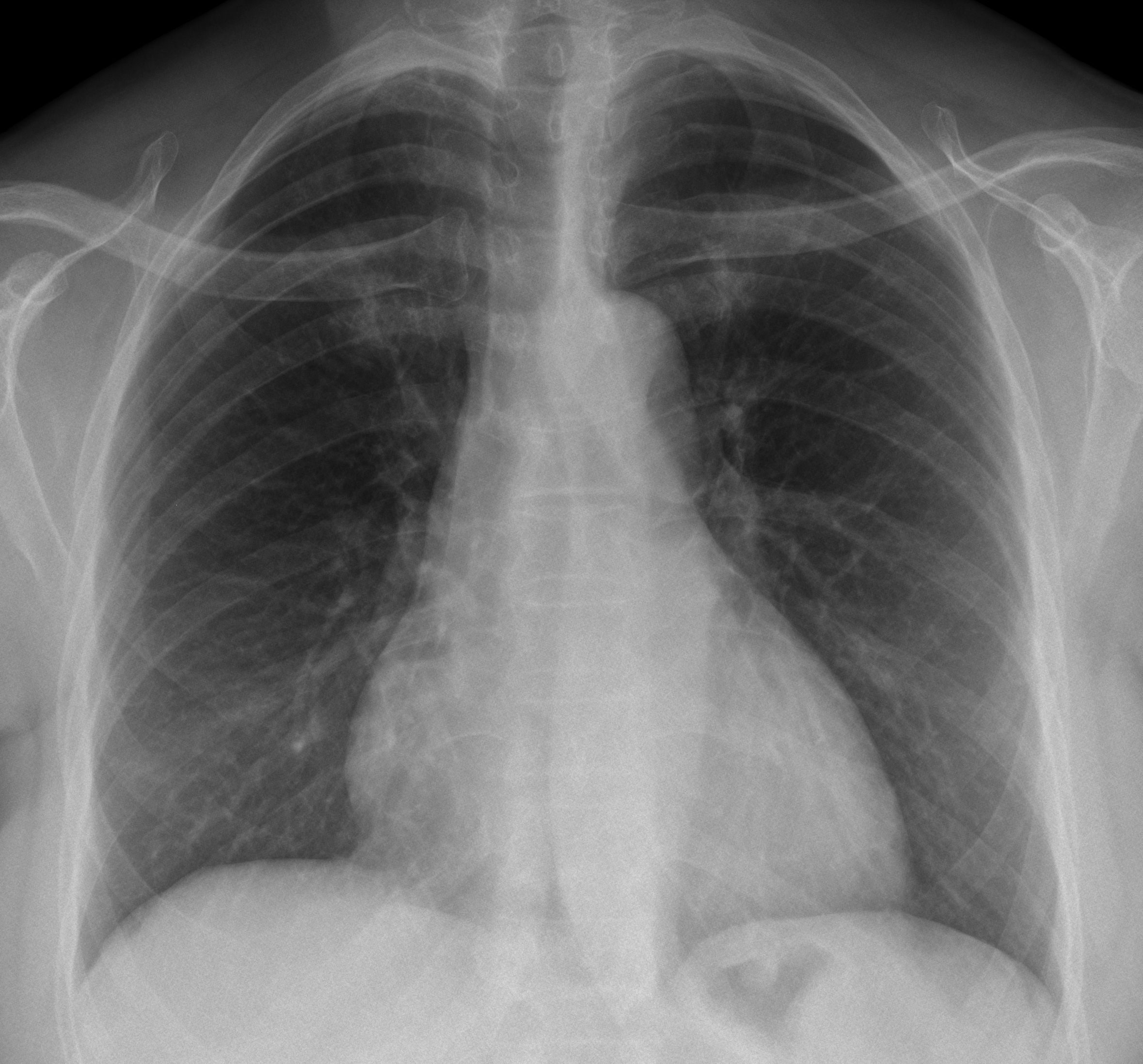
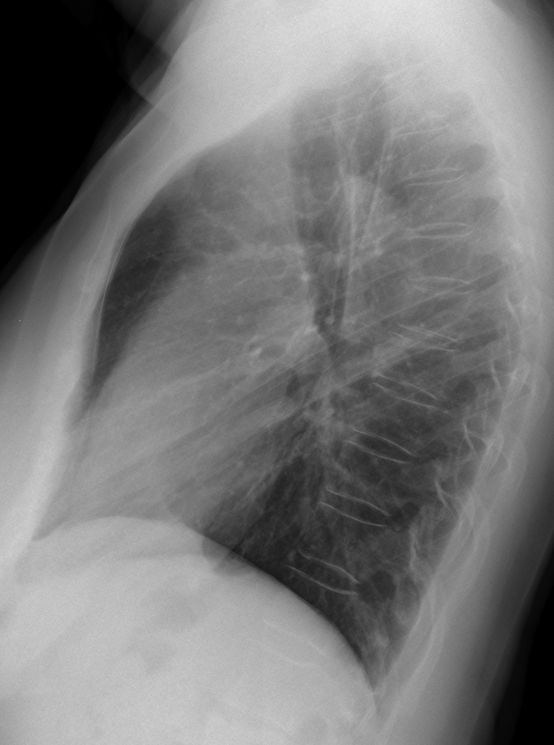
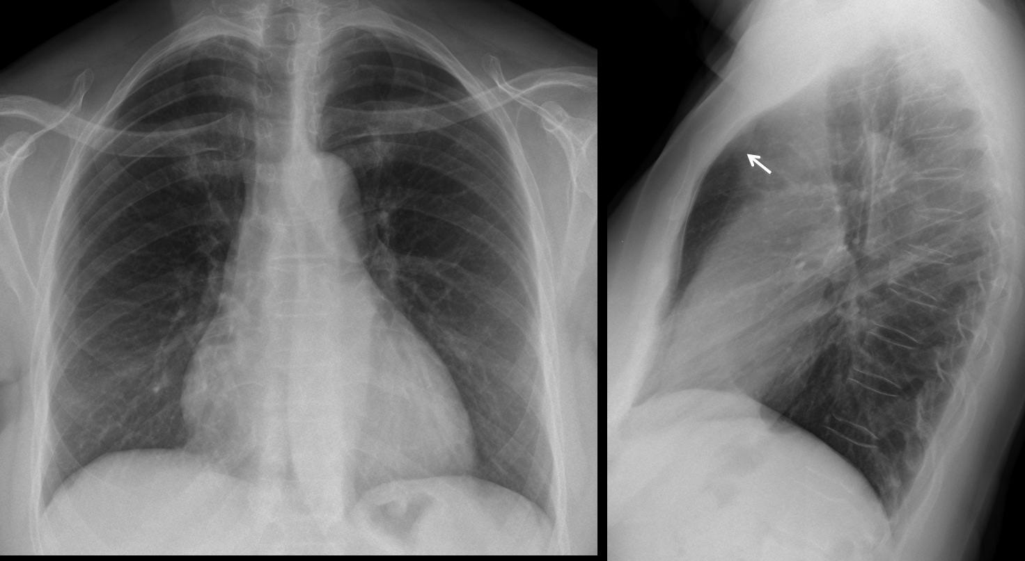
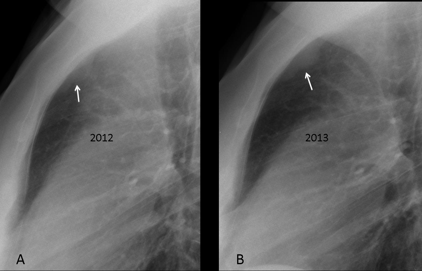
Today we’re presenting a routine chest control of a 53-year-old woman who had a lumpectomy for carcinoma of the breast three years ago. Radiographs were read as normal. Do you agree? Any ideas?
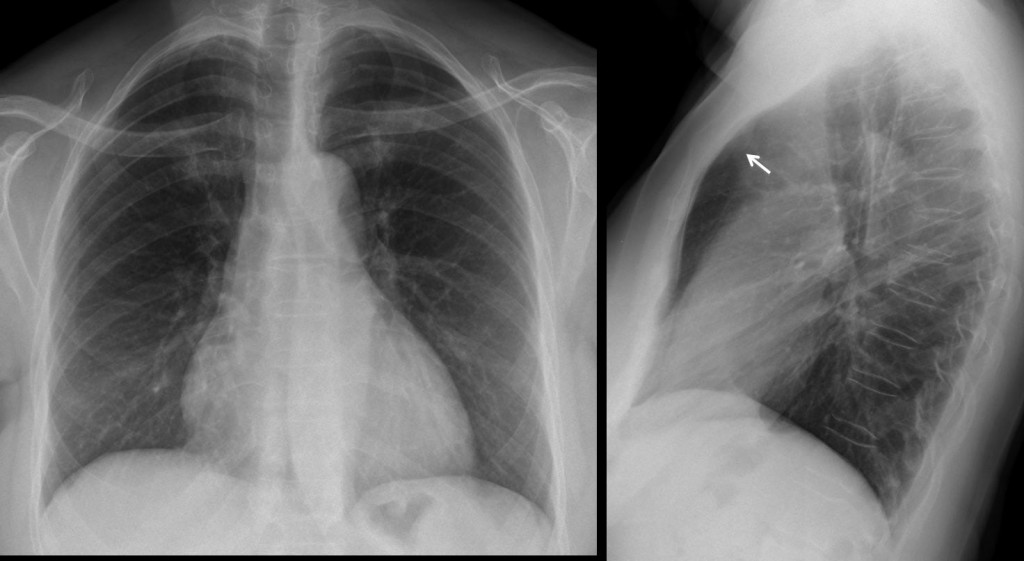
Fig. 1
Findings: the lateral chest radiograph shows a lobulated, well-defined retro-sternal opacity (arrow), better seen in the cone down view (fig. 2a, arrow). The opacity has increased in size one year later (fig. 2b, arrow).
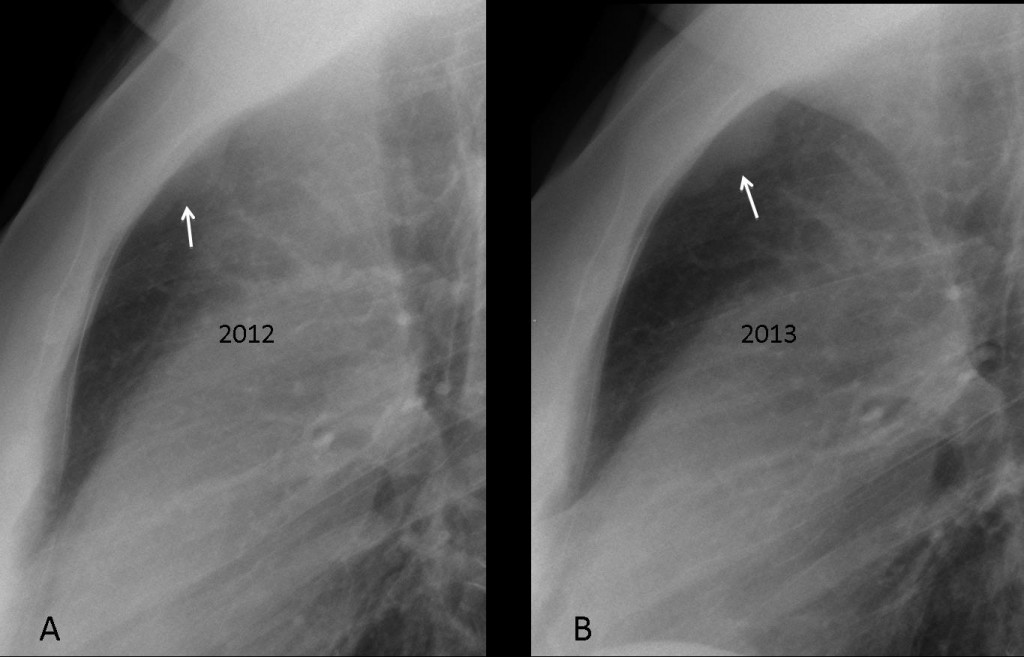
Fig. 2
Sagittal and axial CT confirms the presence of the mass (arrows).
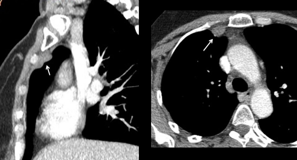
Fig. 3
Final diagnosis: metastases to internal mammary nodes. No other metastases were seen.
I am presenting this case to emphasise the importance of looking in the lateral view at the blind areas in the PA chest radiograph. The metastases were overlooked in the initial exam and were found one year later by somebody more thorough. Congratulations to Katerina, as the sole discoverer of the lesion.
Teaching point: always look at the anterior clear space in the lateral view. Significant findings may be discovered, as in this case.