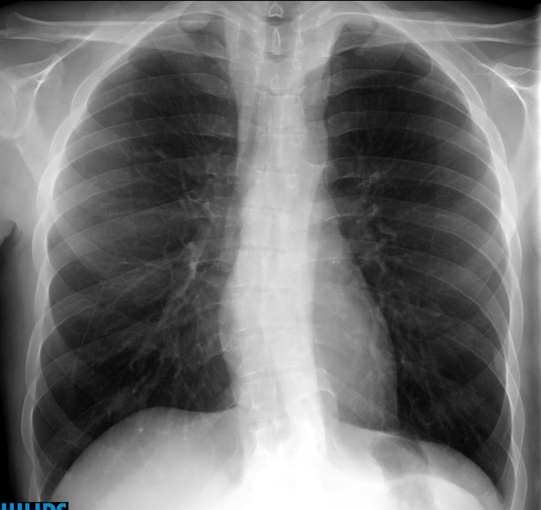
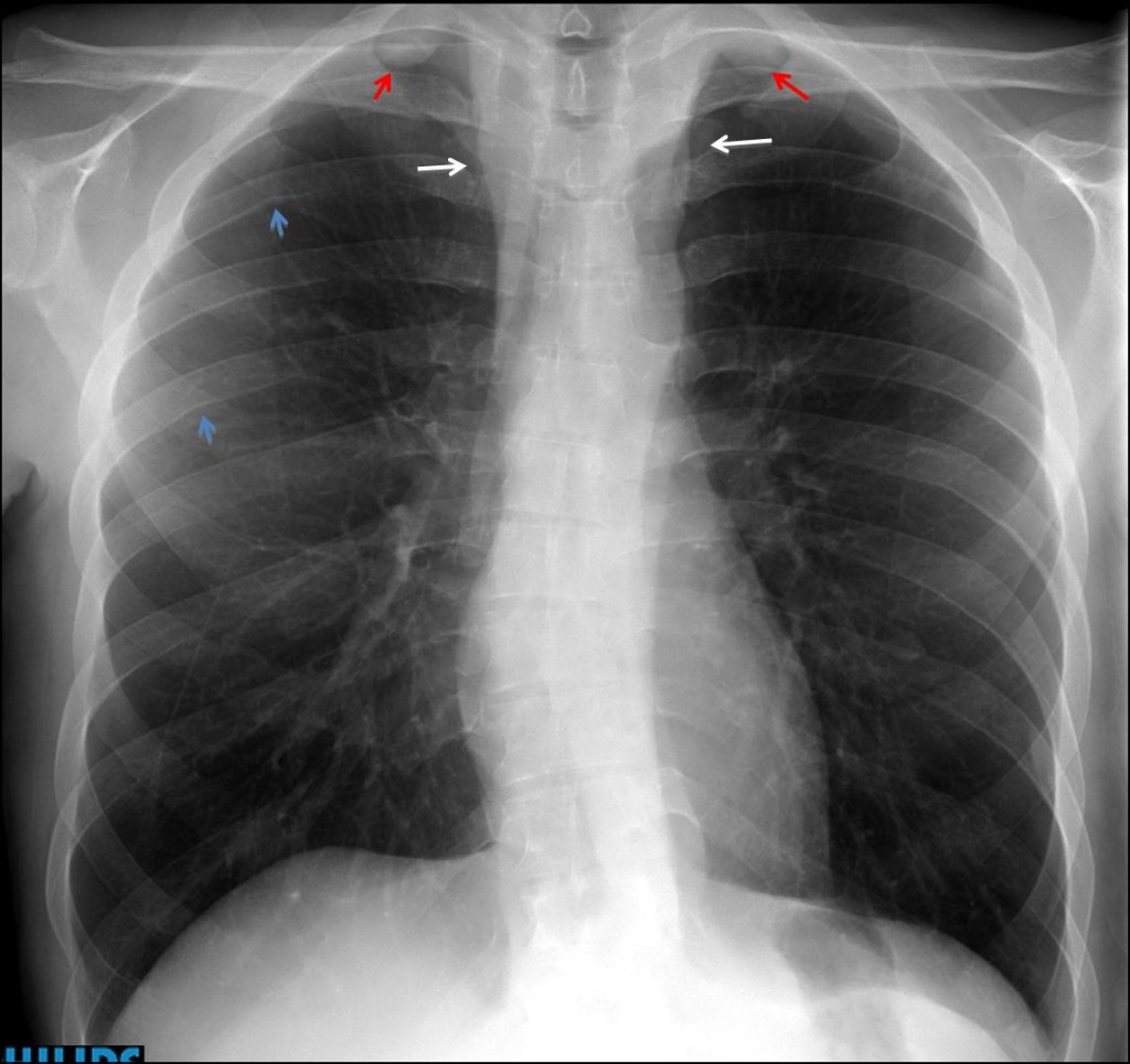
My former disciple Jordi Andreu has contributed this week’s case. Below is a pre-operative PA chest radiograph of a 27-year-old male with a growing perineal mass.
1. SVC obstruction
2. Coarctation of aorta
3. Lymphoma
4. None of the above
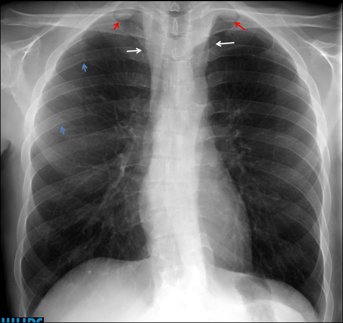
Findings: PA chest shows widening of the superior mediastinum (arrows) and two rounded opacities in both apices (
red arrows). Slight rib notching is visible (
blue arrows). The aortic knob looks normal.
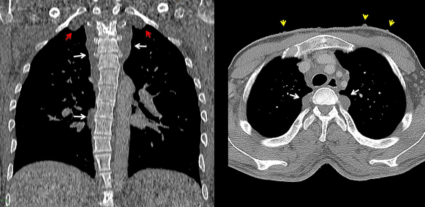
Coronal and axial CT shows soft tissue masses that extend along the posterior mediastinum (arrows) including the extrapleural apical areas (red arrows). The patient had known neurofibromatosis. Multiple skin lesions are visible in the axial CT (yellow arrows).
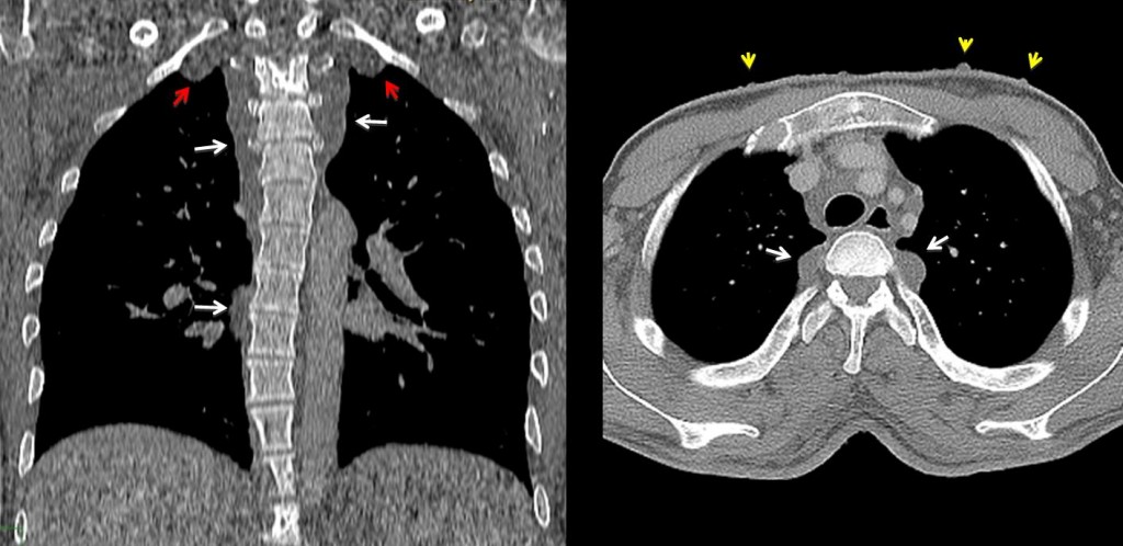
Final diagnosis: neurofibromatosis with sarcomatous degeneration of a perineal lesion.
Congratulations to Katerina, who gave an excellent discussion and made the correct diagnosis.
Teaching point: remember that aortic coarctation is not the only cause of rib notching. Be sure that the patient has hypertension before suggesting coarctation.