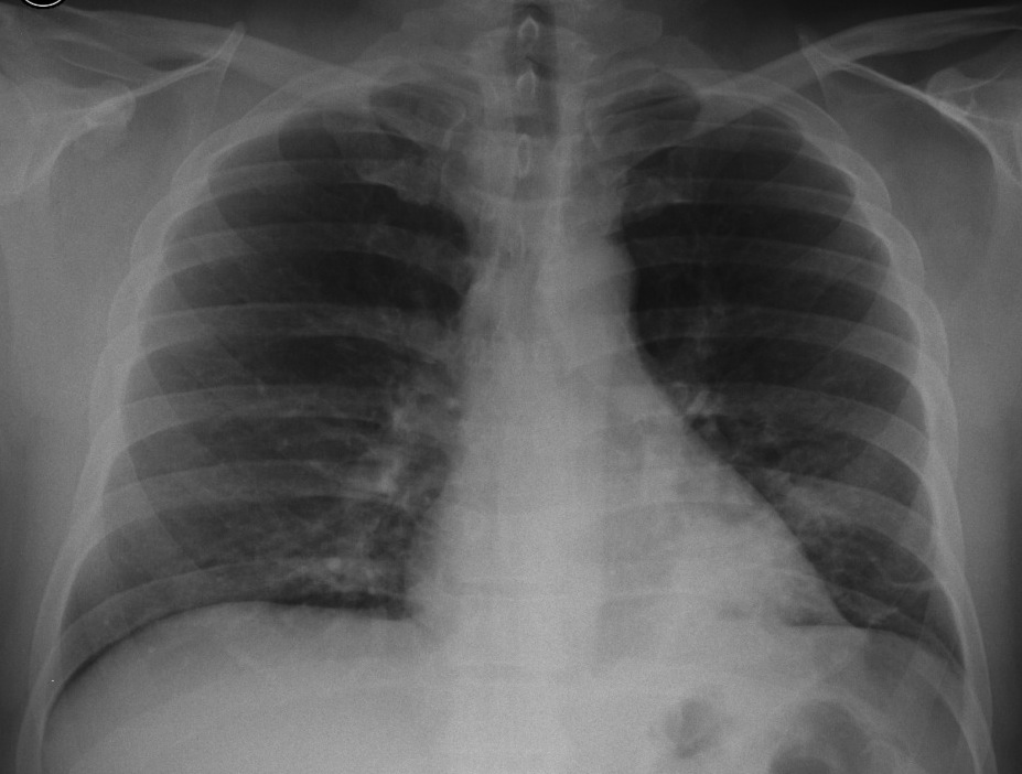
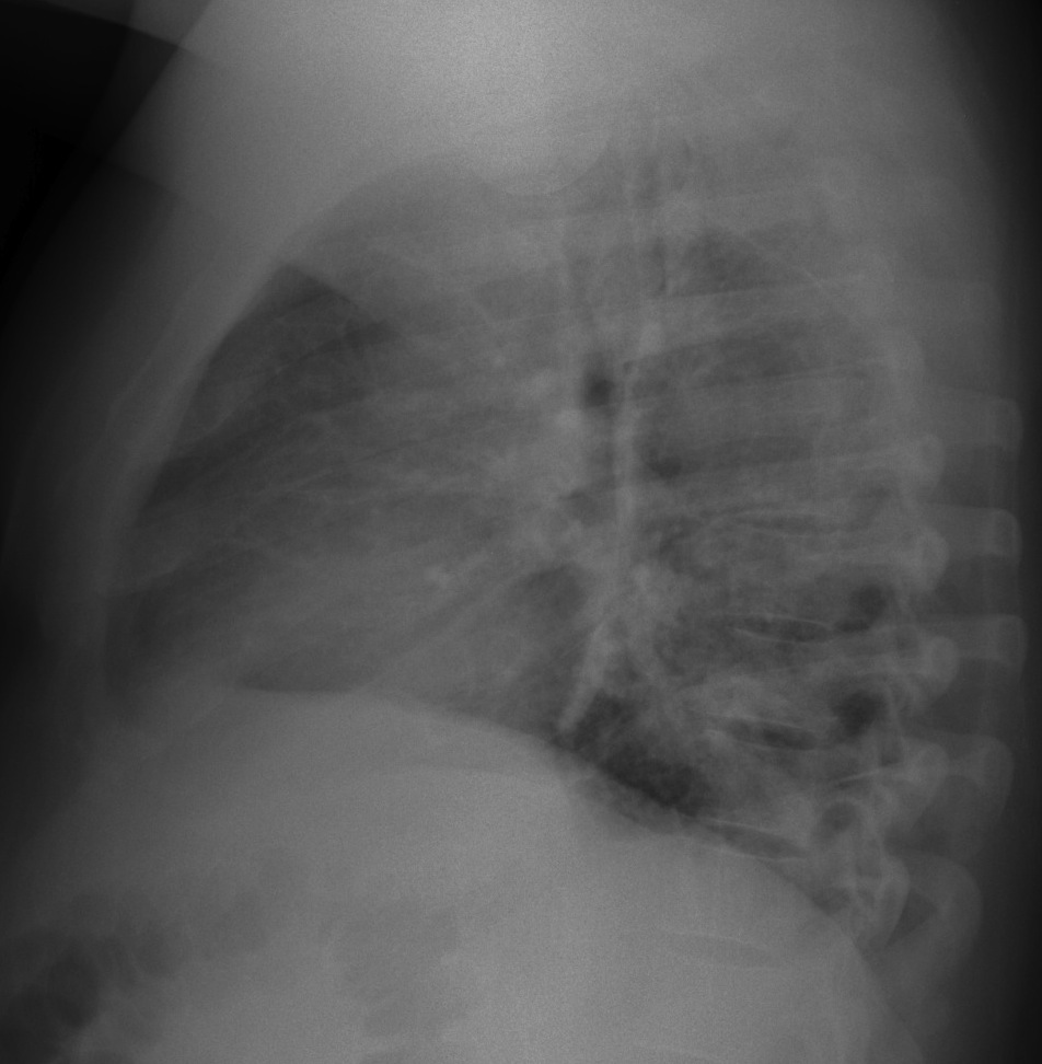
After spending a long weekend with Miss Piggy, Muppet is at peace with the world and has chosen an easy case: 41-year-old male with cough, mild fever and left chest pain.
1. Pneumonia
2. Infected bronchiectasis
3. Pulmonary infarction
4. None of the above
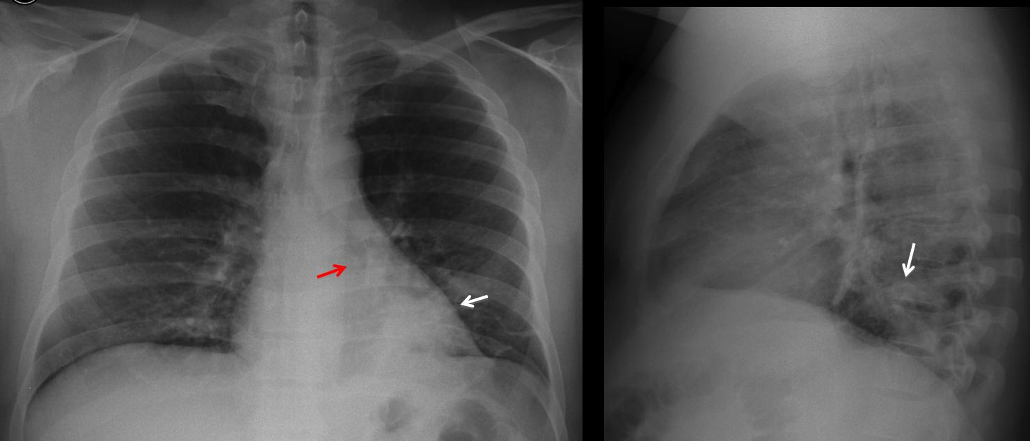
Chest radiographs taken in 2009 show a peripheral opacity in the RLL (arrows). There is marked downward displacement of the left hilum (
red arrow), indicating volume loss.
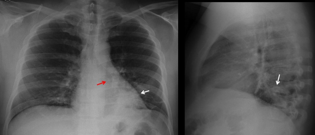
Fig. 1
Hilar displacement was overlooked and the patient did not have new radiographs until February 2013, showing an obvious LLL collapse (Fig 2a, arrows). CT demonstrates an endobronchial lesion (2b, arrow), with fat in the center (2c, arrow). There is marked collapse of LLL (b, c, red arrows). A lobectomy was done.
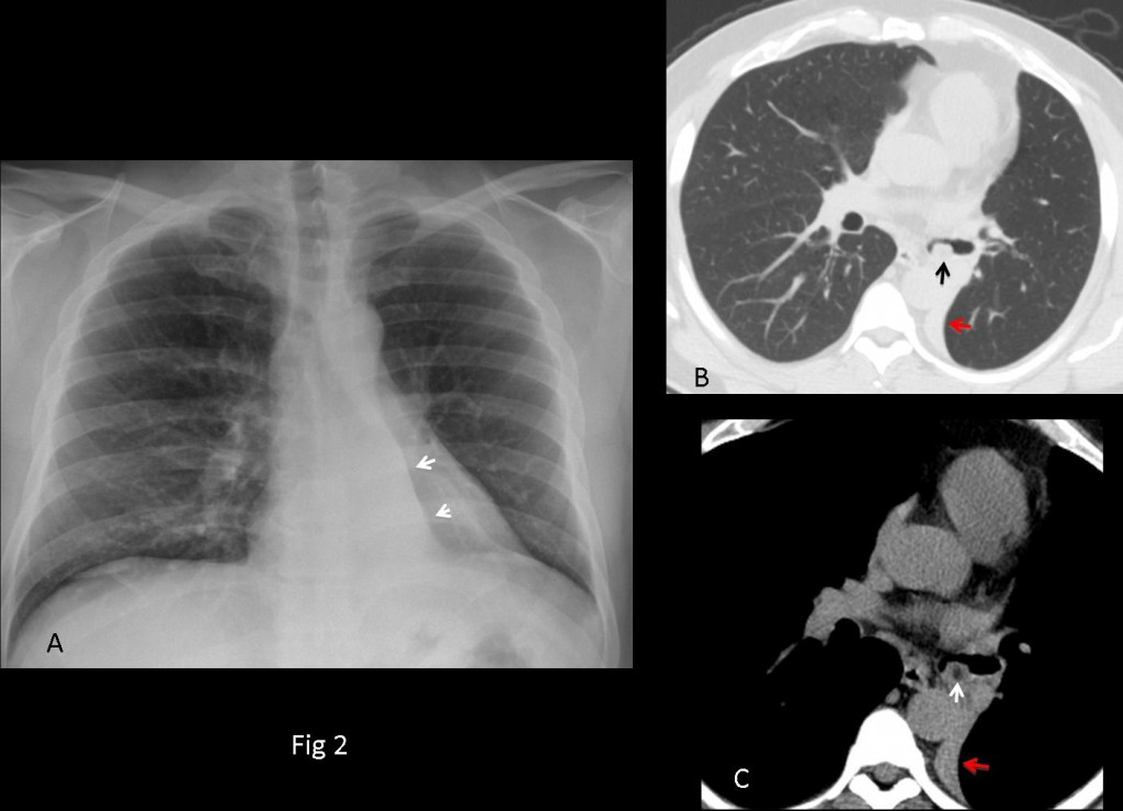
Fig. 2
Final diagnosis: endobronchial hamartoma
Congratulations to Dr. Mapia, who first mentioned the downward displacement of hilum.
Teaching point: don’t forget to look at the hila. In this patient, the diagnosis was delayed four years and it cost him a lobectomy.