Dear Friends,
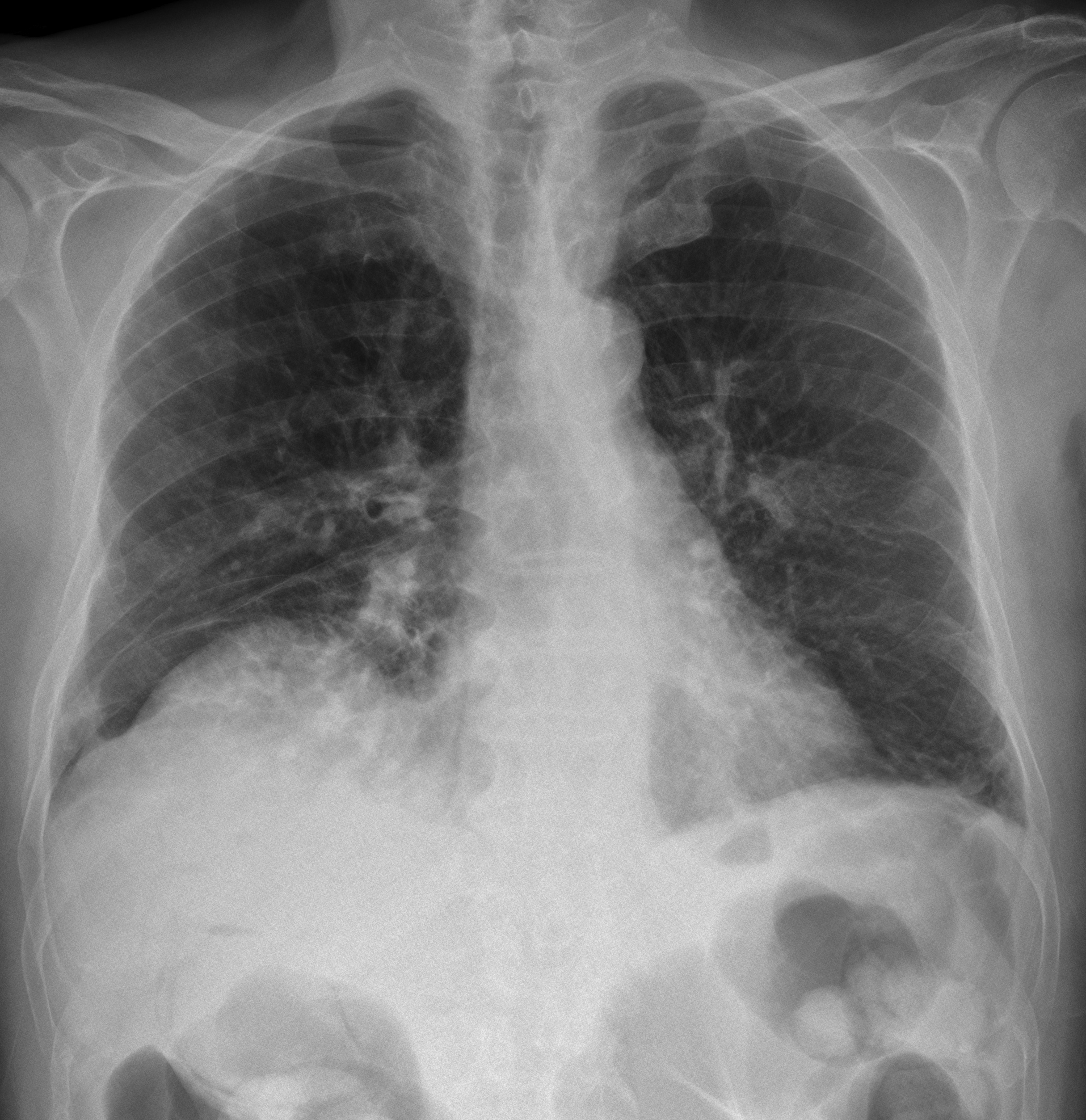
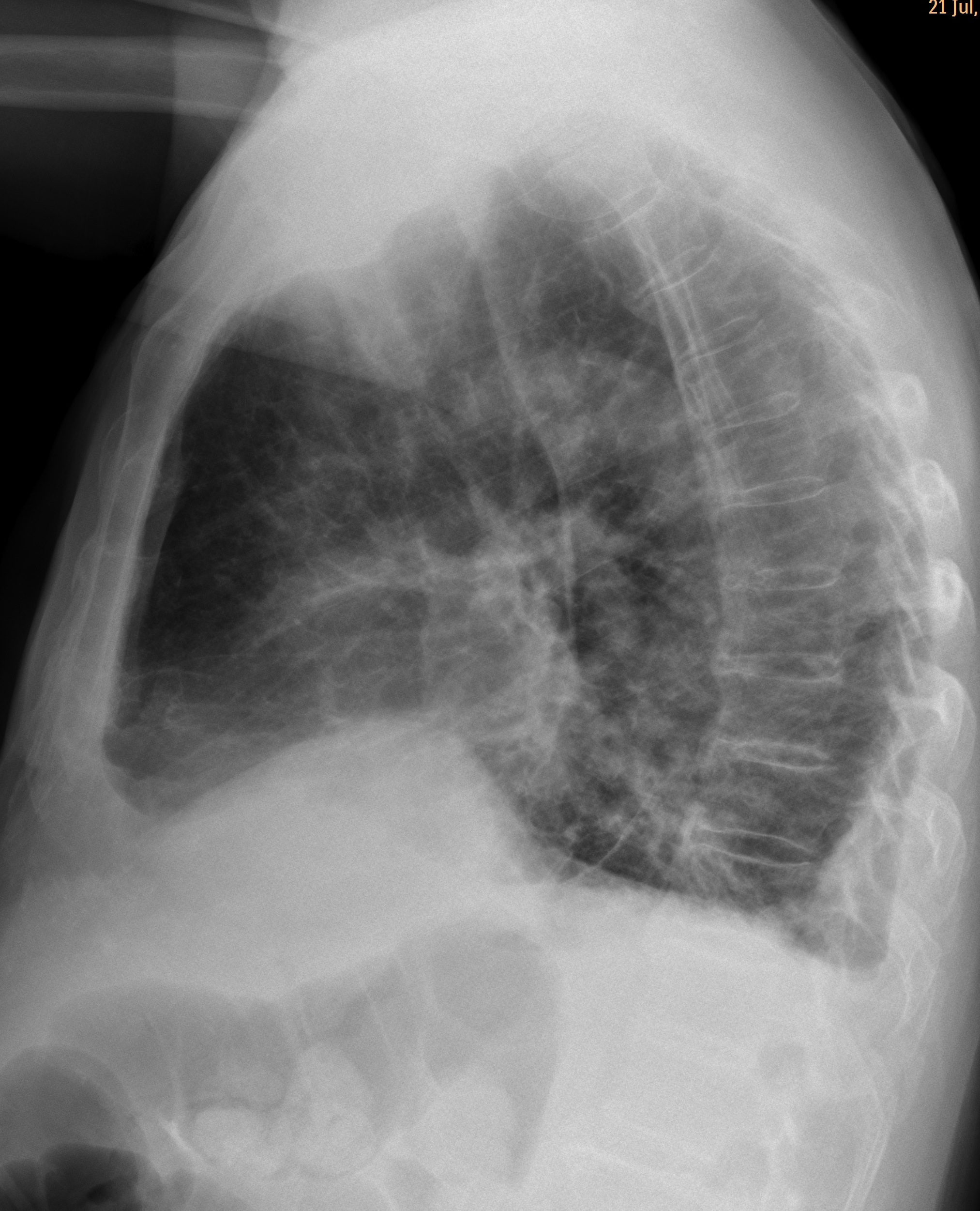
Summer is near and Muppet wants to relax and show easy cases. The present one relates to an 80-year-old male with RUQ pain after embolisation of a liver tumour.
Diagnosis:
1. Pulmonary embolism
2. Pneumonia
3. Subpulmonary fluid
4. None of the above
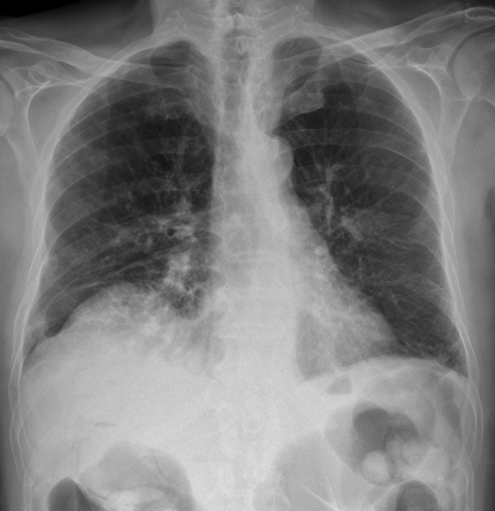
80-year-old male, PA chest
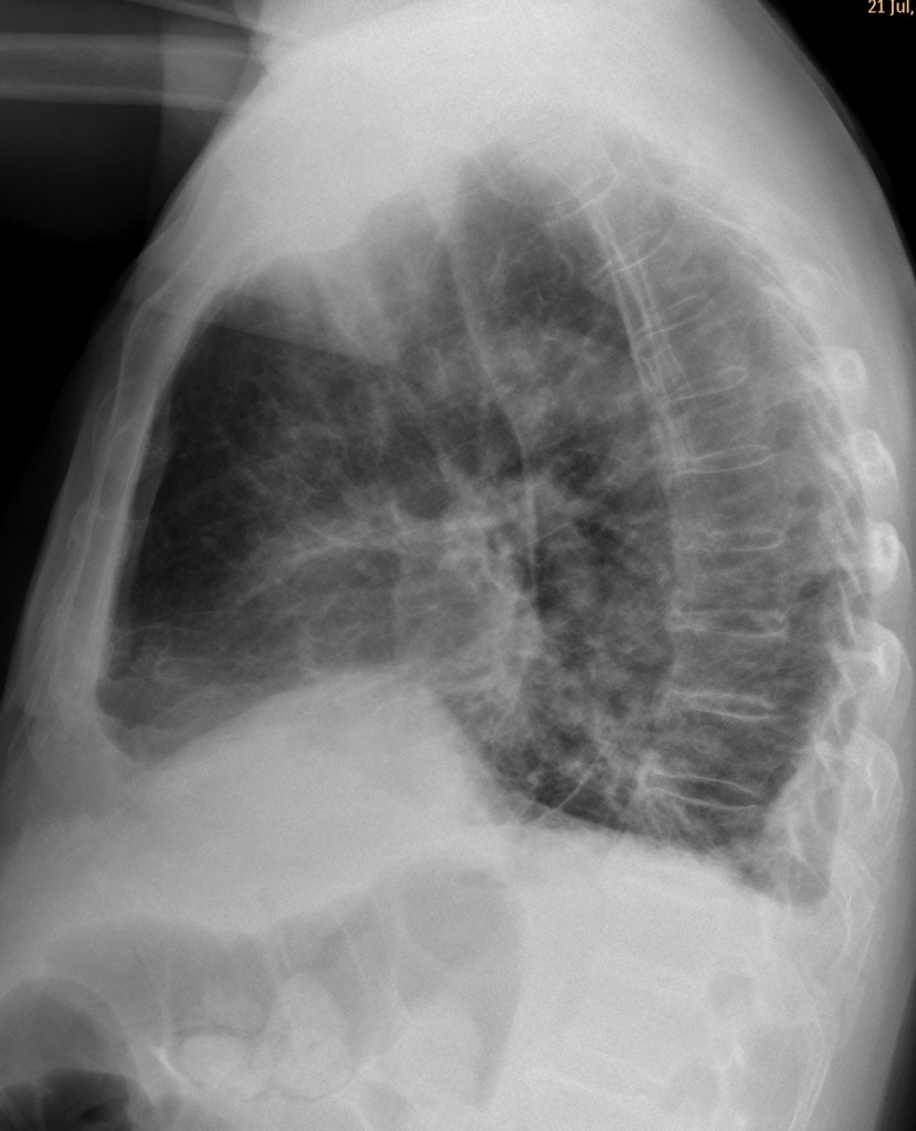
80-year-old male, lateral chest
Leave your thoughts and diagnosis below and look out for the answer next Tuesday.
Click here for the answer to case #68
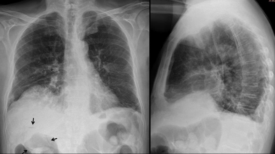
PA chest shows an elevated right hemidiaphragm with ill-defined opacities of the lower right lung. The wall of the gallbladder is outlined by intramural air (arrows). The findings are more obvious in the image that was taken the next day (fig 2 A, arrows).
There is air within the liver (red arrows) and pneumoperitoneum (yellow arrow). CT confirms the intramural gallbladder air (Fig 2 B, arrows) as well as the pneumoperitoneum (B, yellow arrow). The patient died shortly afterwards.
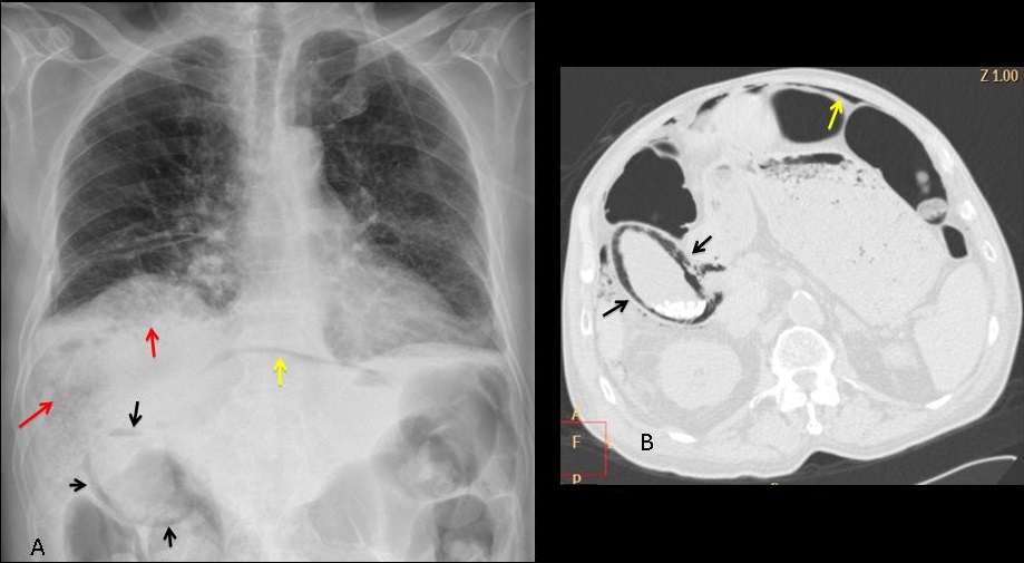
Final diagnosis: gallbladder infarct, secondary to therapeutic embolisation
Congratulations to Dr. Strangelove who made the diagnosis shortly after the case was posted and beat Katerina by 43 minutes.
Teaching point: I was going to stress that you should always look under the diaphragm, but it seems that most of you did.