Dear friends,
Muppet is delighted to present a case provided by Dr. Vilar. The radiographs are of a 57-year-old woman with a chronic cough and mild fever who also suffered from TB in the past.
Diagnosis:
1. TB, bronchogenic spread
2. Hypersensitivity pneumonitis
3. Sarcoid
4. None of the above
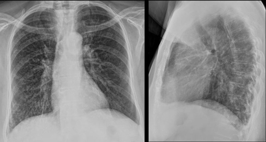
57-year-old woman with a chronic cough and mild fever who also suffered from TB in the past.
Click here for the answer to case #69
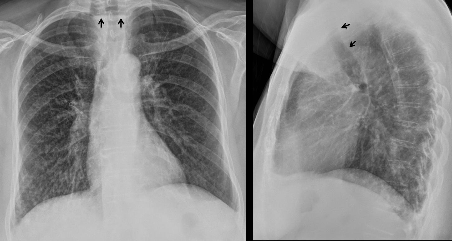
PA chest shows elevation of left hilum, secondary to old TB. There is a diffuse small nodular infiltrate more evident in the right lung, especially in the RLL. An air-fluid level is visible in the upper mediastinum (arrows). The lateral view shows anterior displacement of the trachea (arrows) by a retrotracheal mass.
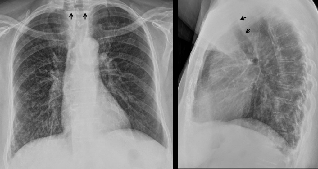
The findings are highly suggestive of a dilated upper oesophagus or a Zenker’s diverticulum with lung aspiration. Next step is an oesophagogram, which shows a large Zenker’s diverticulum (arrows), with aspirated barium in the trachea (red arrows).
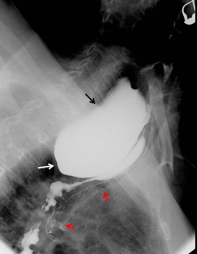
Final diagnosis: Zenker’s diverticulum with bronchial aspiration.
Congratulations to Katerina, who gave an excellent discussion and suggested the right diagnosis.
Teaching point: pulmonary infiltrates in the right lung or RLL should raise the possibility of aspiration. An oesophagogram is an easy procedure to confirm or discard the diagnosis.