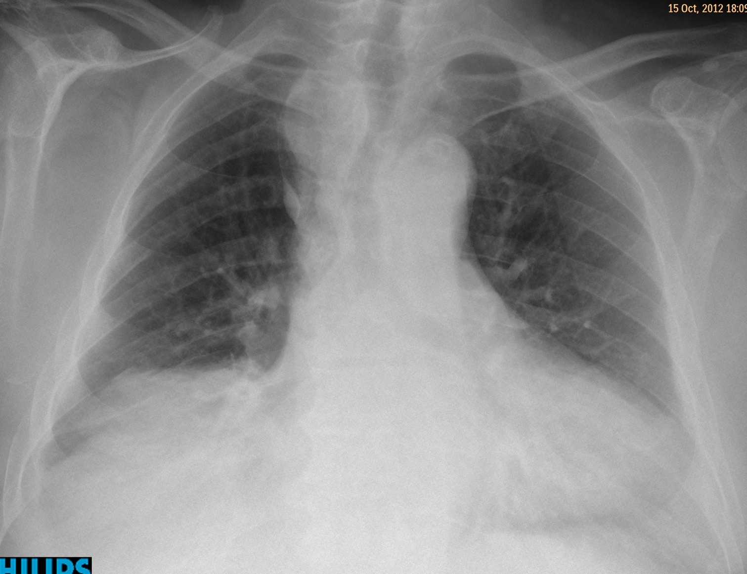
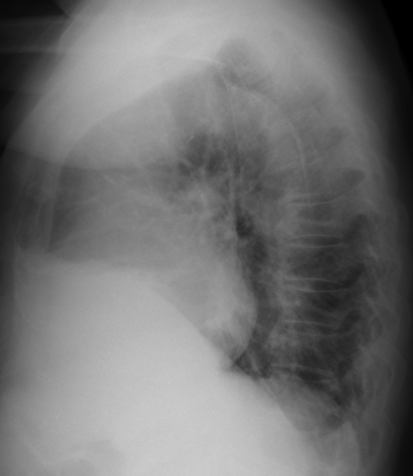
Showing images of a pre-operative chest for inguinal hernia in a 72-year-old male.
1. Thyroid mass
2. Thymoma
3. Lymphoma
4. None of the above
Findings: PA radiograph shows an apparent right upper mediastinal mass (white arrow), slightly displacing the trachea. There is an associated azygos lobe (
red arrow). Lateral view is non-contributory.
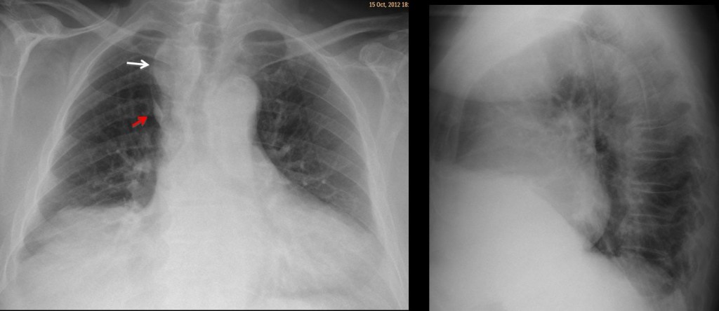
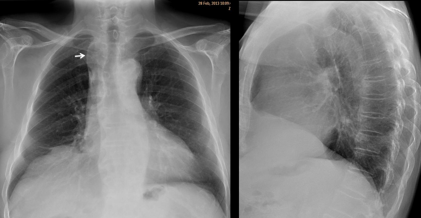
Findings were interpreted as a normal variant, the opaque azygos lobe, in which tortuous supra-aortic vessels in combination with an azygos lobe give the impression of a right upper mediastinal mass. In this particular case, films were not taken in full inspiration, which increased the false impression of a mass. Radiographs taken five months later confirm the original diagnosis (Fig 2).
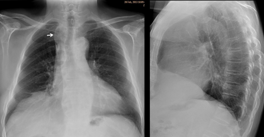
Fig 2. Note the opaque azygos lobe (arrow)
Final diagnosis: opaque azygos lobe (ref. AJR 160:245-48, 1993)
Congratulations to Katerina who reconsidered her original impression and suggested a vascular lesion.
Teaching point: forgive my insistence but, as stated in case 42, remember that vessels compose the mediastinum, basically.