Dr. Pepe’s Diploma Casebook: Case 113 – SOLVED!

Dear Friends,
Glad to be back. I have missed my fans! Planning big surprises for next year. In the meantime, have a look at this preoperative chest radiograph for goiter in a 47-year-old woman. What do you see?
Check the image below, leave your thoughts in the comments section and come back on Friday for the answer.

Findings: PA radiograph shows a barely visible cystic image at the cardiophrenic angle (A, black arrows), better seen in the coned-down view (B, black arrows). An air-fluid level is visible at the bottom of the cavity (A and B, red arrow).
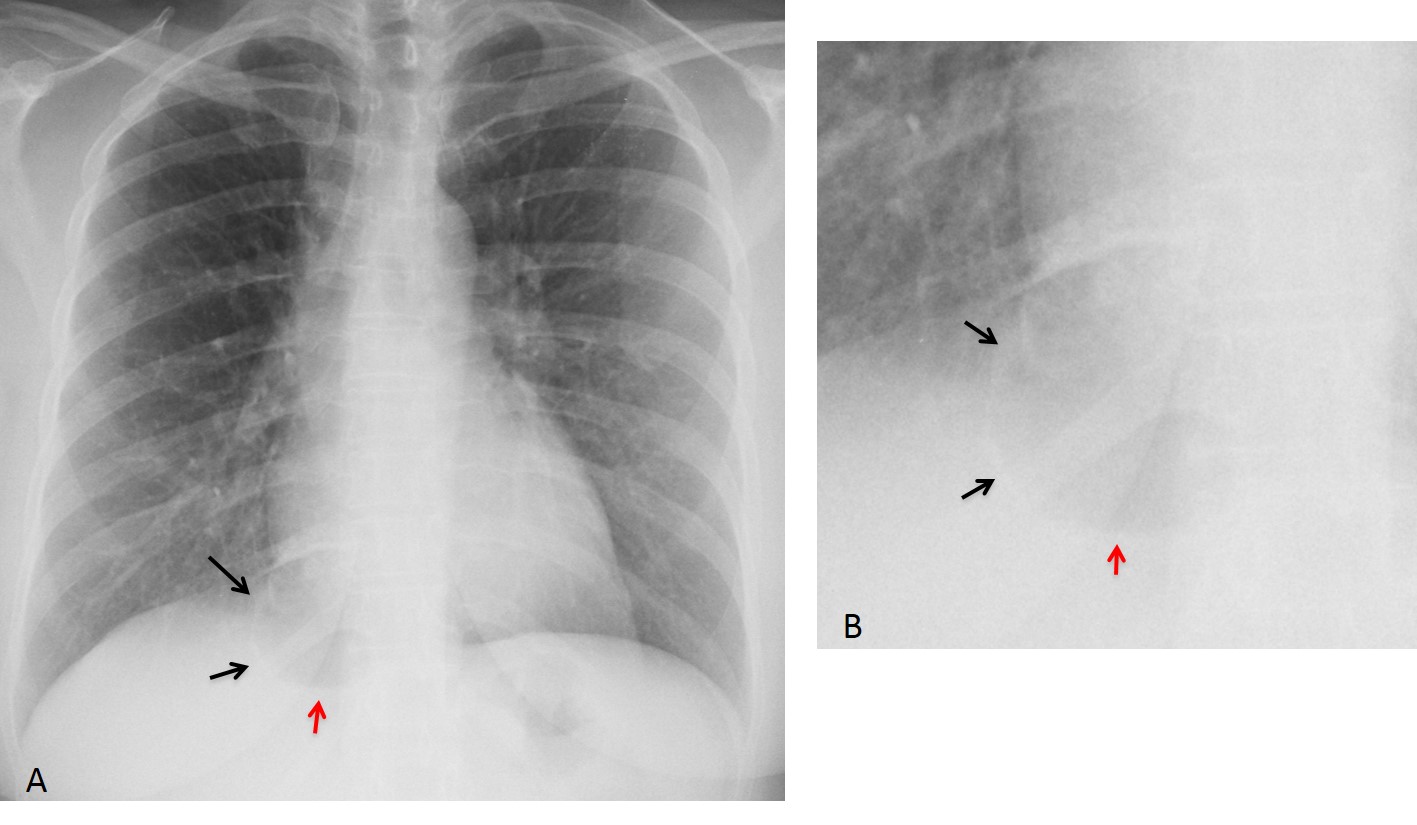
Enhanced CT shows a multicystic lesion in the medial aspect of the RLL (C and D, arrows), with an air-fluid level in the larger cyst (E, red arrow). No systemic artery is visible.
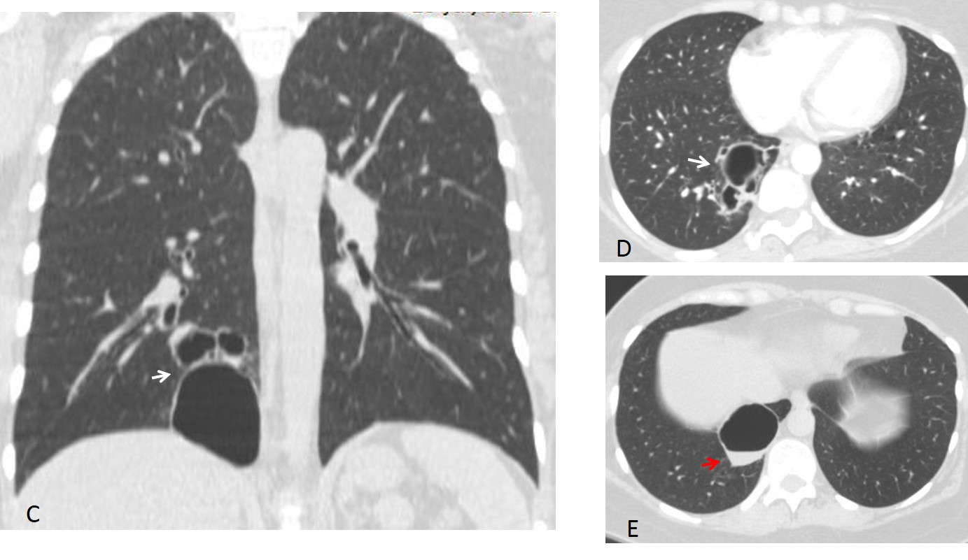
Final diagnosis: congenital cystic malformation of the lung.
I am showing this case to discuss the right paracardiac space, an area that includes the medial portion of the right lower lobe, the lower right mediastinum, and the medial right hemidiaphragm (Fig. 1). Unless we specifically focus our attention on this area, subtle abnormalities may be overlooked.
Disease in this area may arise from the lung, lower mediastinum, or diaphragm.
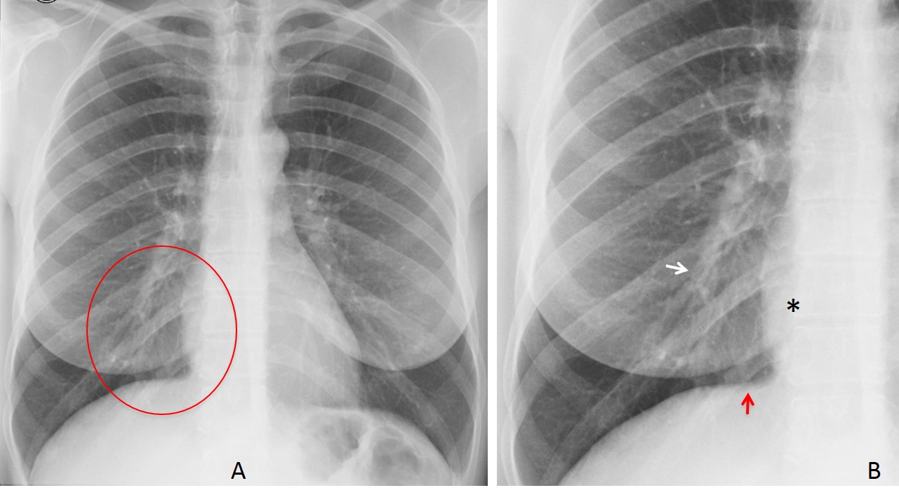
Fig. 1
Fig. 1. Normal chest. The oval in A depicts the right paracardiac space. The components are better seen in the magnified view: lower lobe vessels (B, white arrow), lower mediastinum (B, asterisk), and diaphragm (B, red arrow).
Lung pathology in this area can be obscured by the right lower lobe vessels. Any lung condition can occur in the right paracardiac space. The most common is pneumonia (Fig. 2), although other pathologies may occur as well (Fig. 3).
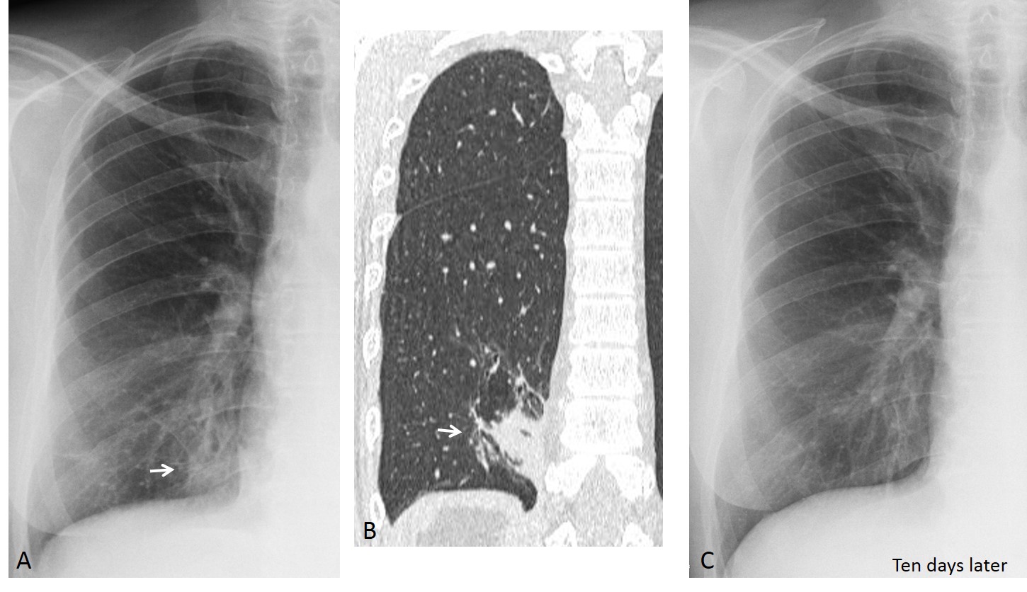
Fig. 2
Fig. 2. Poorly visible pneumonia at the right paracardiac space (A, arrow), confirmed with CT (B, arrow). Ten days later, the pneumonia has disappeared (C).
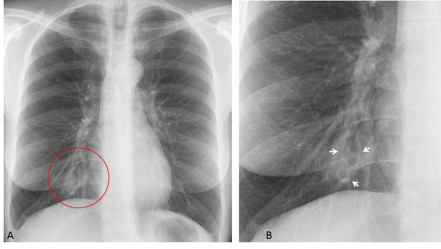
Fig. 3
Fig. 3. Poorly-defined RLL opacity in an asymptomatic patient (A, circle). Coned-down view shows tortuous vessels (B, arrows), interpreted as a possible AV malformation. Unproven.
Lung nodules located in the right paracardiac space may be missed, either because they are obscured by adjacent structures (Fig. 4) or because of their small size (Fig. 5).

Fig. 4
Fig. 4. Nodular lesion at the right cardiophrenic angle (A, white arrow). Coarse calcification is detected within (A, red arrow) and confirmed by unenhanced coronal CT (B, arrow). Diagnosis: lung hamartoma.
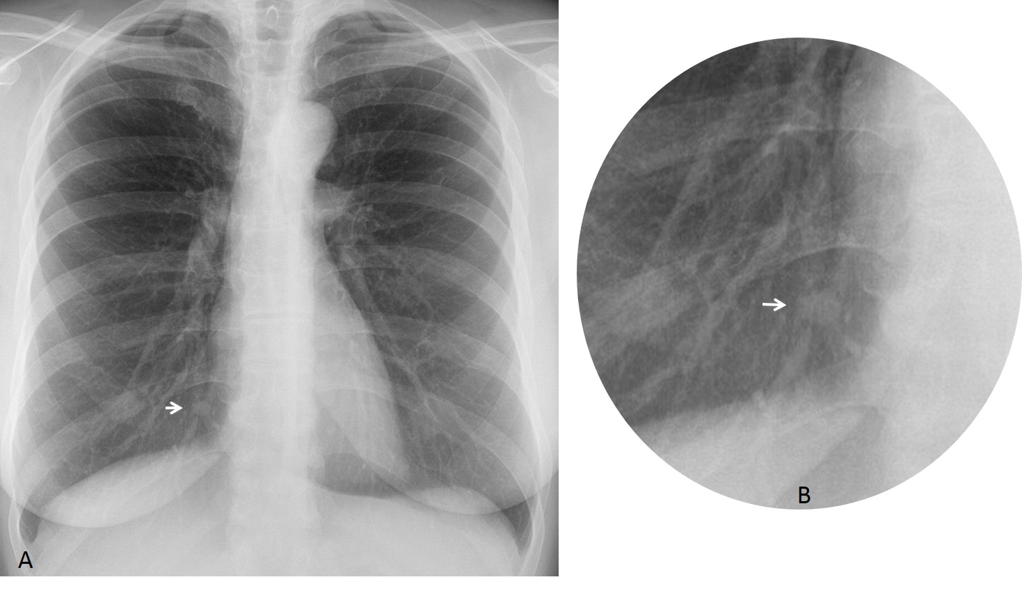
Fig. 5
Fig. 5. Preoperative chest radiograph in a 42 y.o. woman. A small paracardiac nodule was seen (A and B, arrows). PET was positive and surgery was performed. Diagnosis: benign spindle-cell tumor.
Lower mediastinal masses may be overlapped by the cardiac shadow. They can be suspected on detection of increased density of the right cardiac silhouette compared to the left side (Fig. 6) or because they give a double contour to the right lower mediastinum (Figs. 6 and 7).
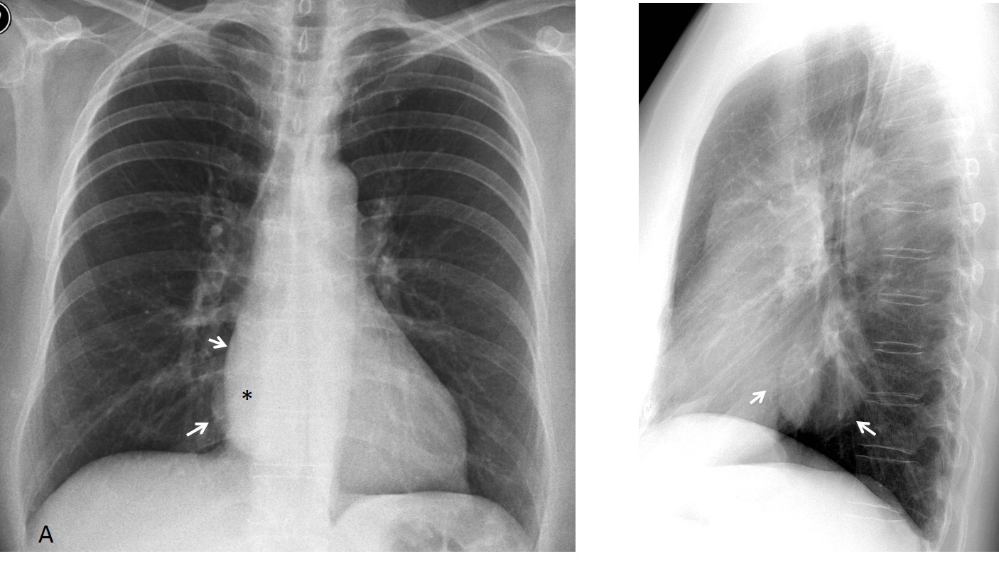
Fig. 6
Fig. 6. Preoperative radiograph shows a double contour of the right lower mediastinum (A, arrows). In addition, the right side of the heart (A, asterisk) is denser that the left side. The lateral view shows a well-defined middle mediastinal mass (B, arrows). Review of films taken six years earlier show the mass unchanged. Diagnosis: duplication cyst.
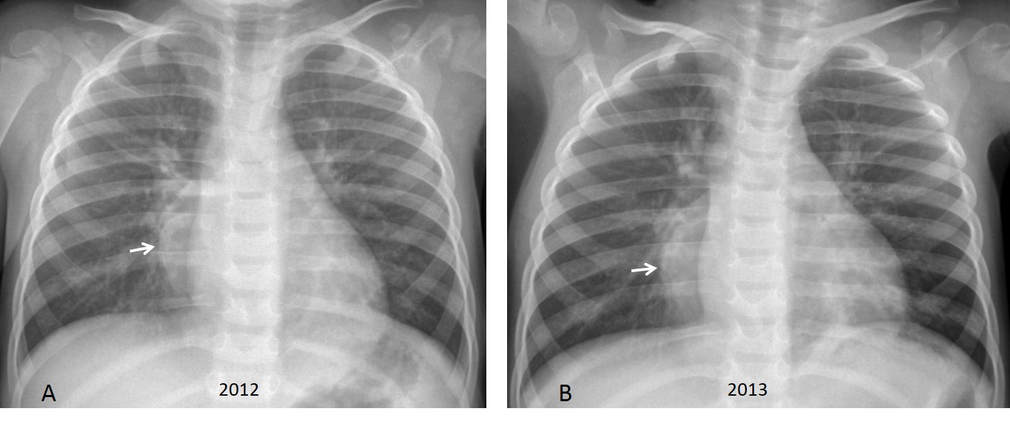
Fig. 7
Fig. 7. Seven y.o. boy with a history of pneumonia in two consecutive years. A right posterior mediastinal mass was missed in both examinations, despite the obvious double contour of the right lower mediastinum (A and B, arrows).
Three years later the mass has grown considerably (C and D, arrows). Diagnosis: neuroblastoma.
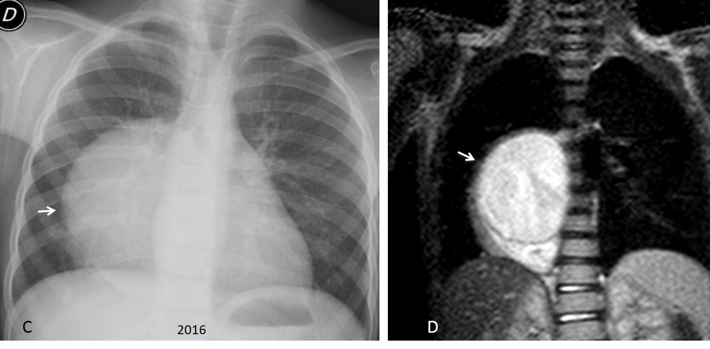
Fig. 7
Lesions arising from the diaphragm are usually Morgagni hernias, which appear as masses at the cardiophrenic angle. The main component is abdominal fat (Fig. 8). Sometimes air is visible when fat is accompanied by herniated bowel (Fig. 9).
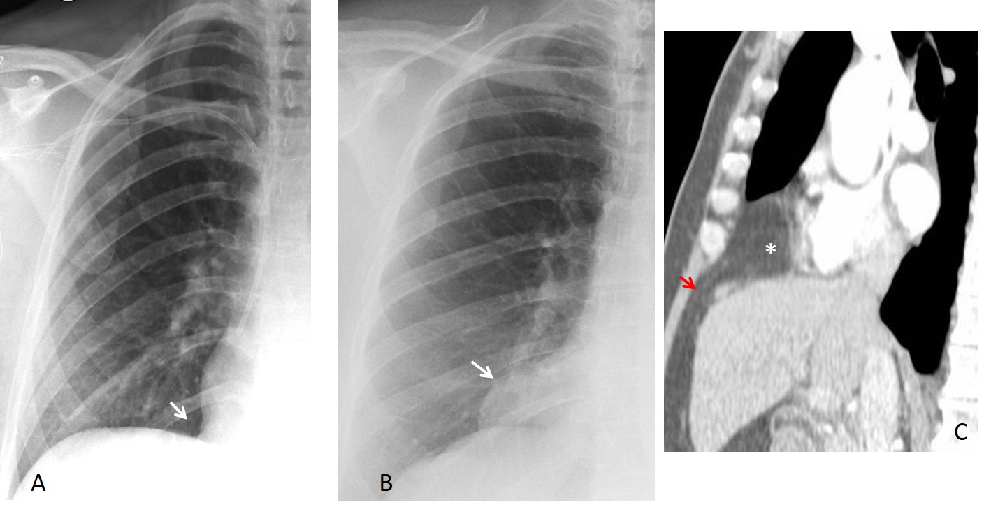
Fig. 8
Fig. 8. Morgagni hernia after abdominal surgery. Preoperative radiograph shows a normal cardiophrenic angle (A, arrow). One year later a mass has appeared in the same location (B, arrow). Enhanced sagittal CT shows herniated abdominal fat (C, asterisk) through a hiatus in the diaphragm. Note the vessels crossing the hiatus (C, red arrow).
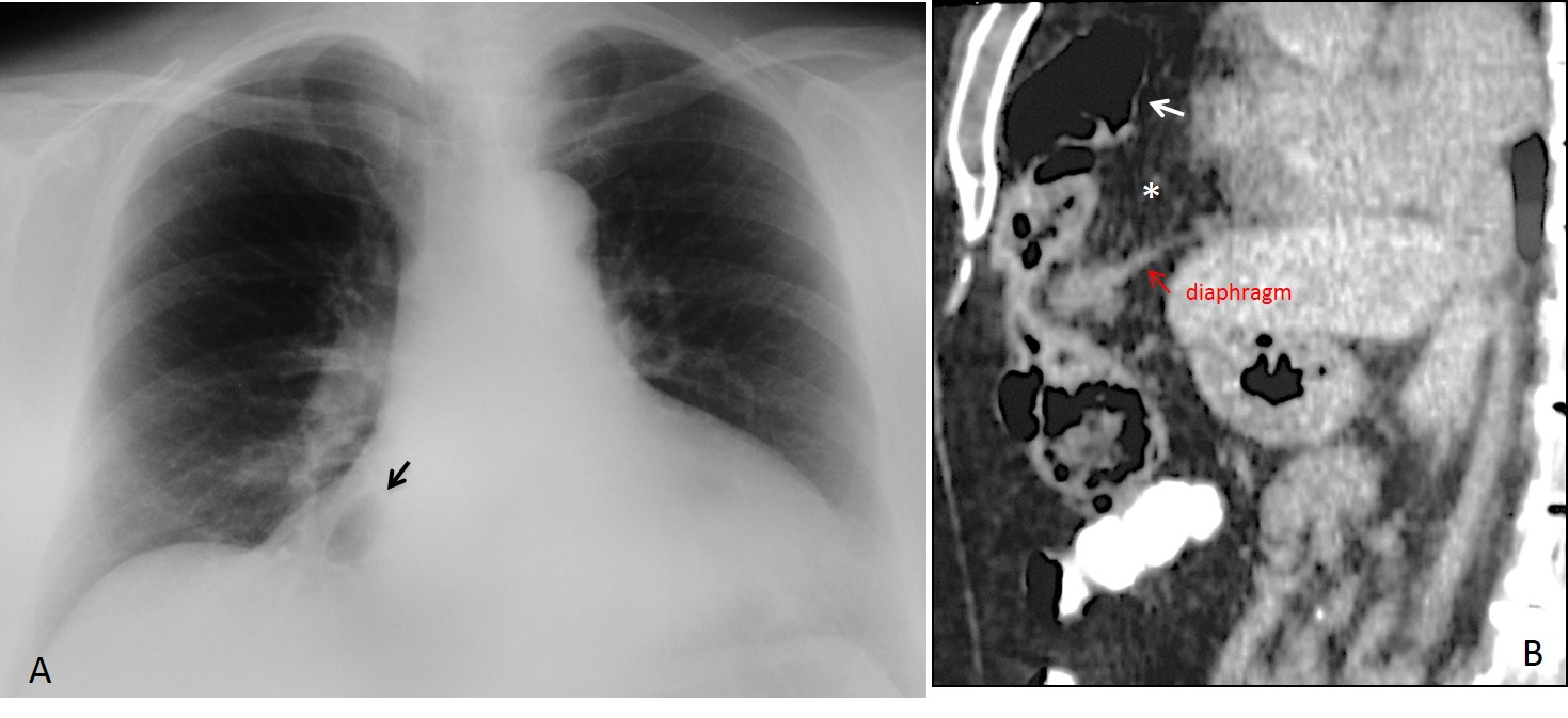
Fig. 9
Fig. 9. Morgagni hernia after abdominal surgery. Preoperative radiograph shows a normal cardiophrenic angle (A, arrow). One year later a mass has appeared in the same location (B, arrow). Enhanced sagittal CT shows herniated abdominal fat (C, asterisk) through a hiatus in the diaphragm. Note the vessels crossing the hiatus (C, red arrow).
Looking at the right paracardiac space can be very rewarding, as is shown in the case below, in which an unsuspected intrathoracic rib was discovered (Fig. 10) after having been missed in previous examinations.
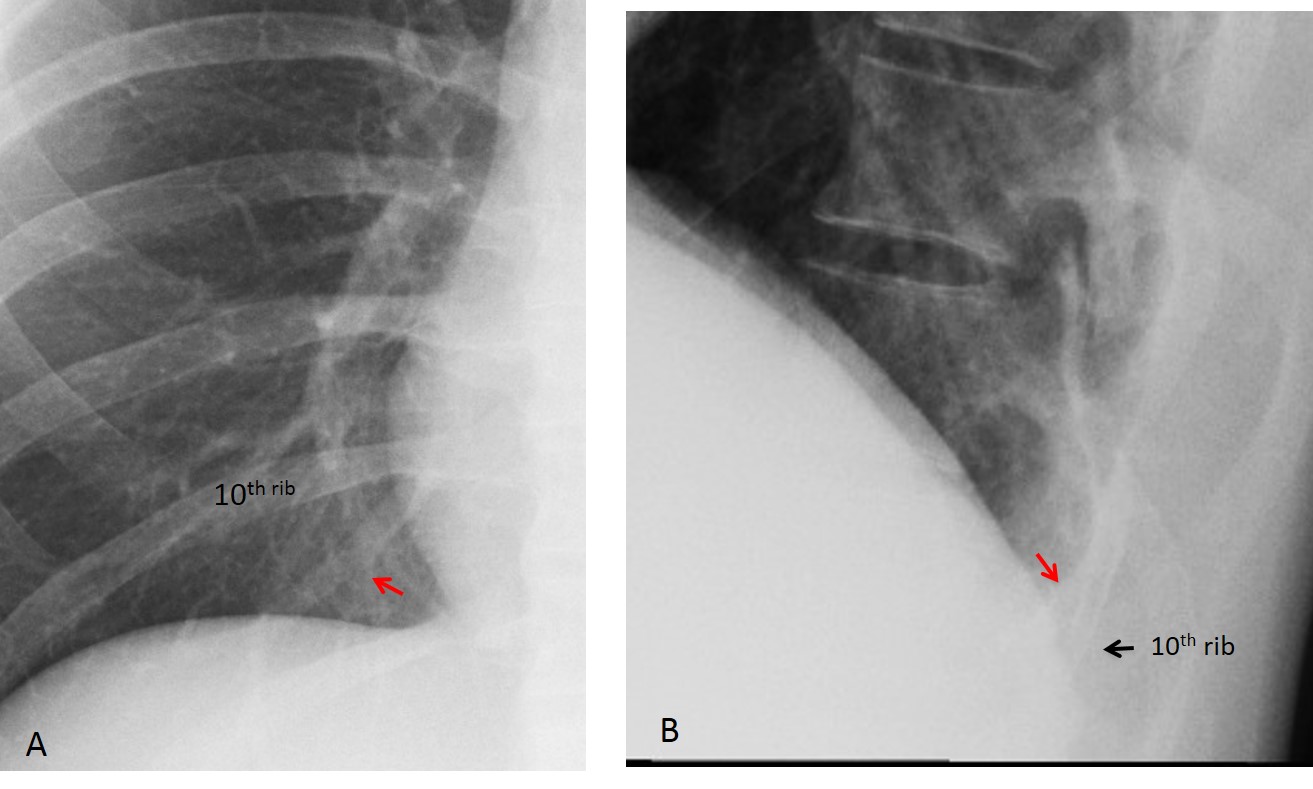
Fig. 10
Fig. 10. Preoperative chest radiograph showing an intrathoracic rib as an incidental finding (A and B, red arrows). The anomalous rib is connected to the tenth right rib.

Follow Dr. Pepe’s advice:
1. Lesions in the right paracardiac space are often overlooked unless we specifically focus our attention on this area.
2. Pathology in the right paracardiac space can arise from the lung (infiltrates, nodules), lower mediastinum (masses), or diaphragm (Morgagni hernia).


