Dr. Pepe’s Diploma Casebook: Case 116 – SOLVED!
Dear Friends,
This will be the last Diploma Casebook case posted on the ESR Blog. Following the steps of Neymar, I have been offered a succulent contract by the European Board of Radiology and next year I will posting my cases on the EBR website, starting on Monday, January 15. I am excited about this new challenge and hope you will be too!
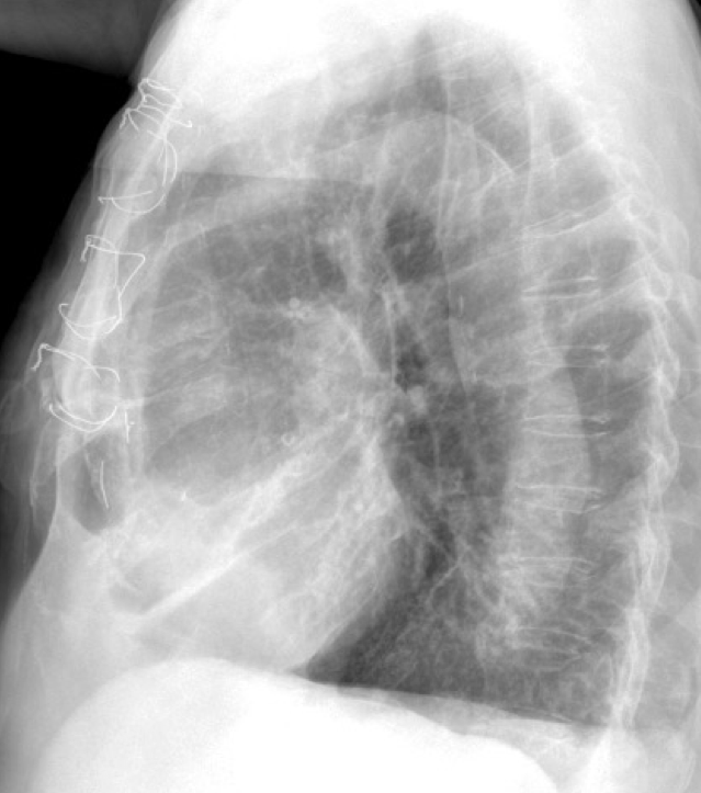
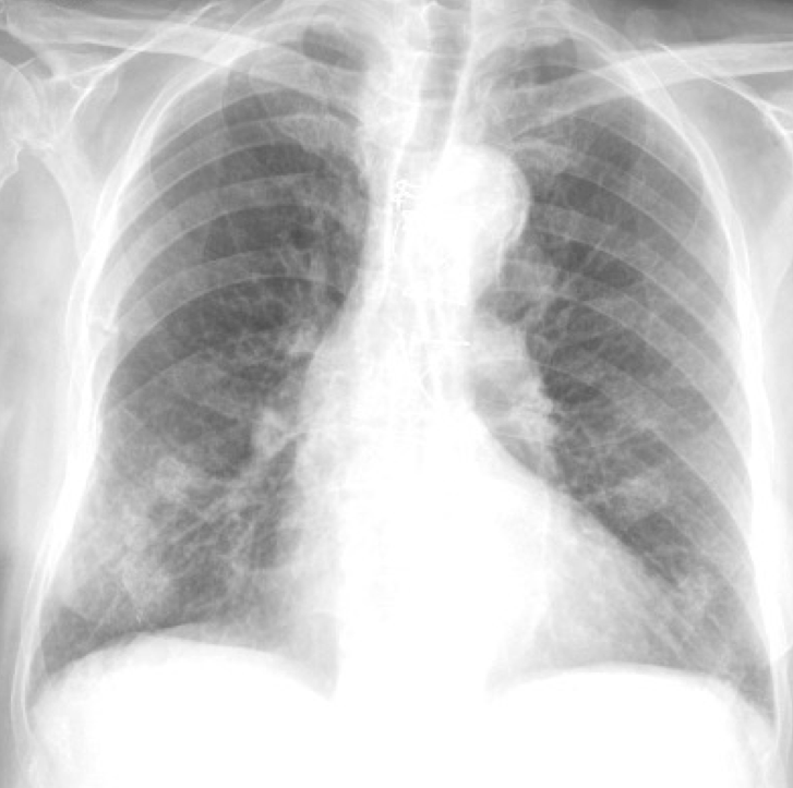
Radiographs of today’s case belong to a 76-year-old man with pain in the chest after a fall. What do you see?
Check the images below, leave your thoughts in the comments section, and come back on Friday for the answer.
Findings: PA radiograph shows numerous rib fractures (A, white arrows), some of which simulate pulmonary nodules (A, yellow arrows). An ancillary finding is a mediastinal opacity at the aorto-pulmonary window (A, red arrow), also visible in the lateral view (B, red arrow).
Comparison with previous films shows progression of the opacity over a three-year period (arrows).
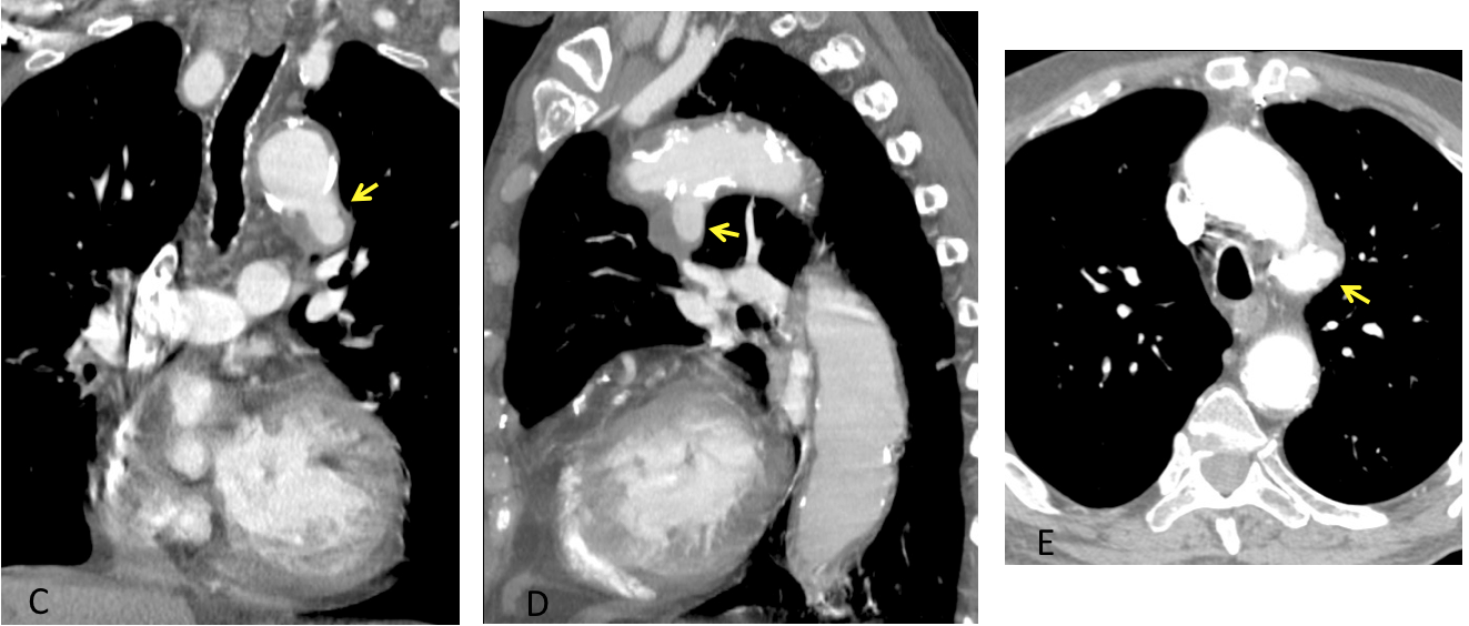
Enhanced CT confirms a partially thrombosed aneurysm arising from the inferior aspect of the aortic arch (C-D and E, arrows)
Final diagnosis: Slow-growing aneurysm of the aortic arch
(Case courtesy of José Vilar, MD)
I am presenting this case to discuss changes in the aortic arch, which is visible as the aortic knob in the PA chest radiograph. From the diagnostic viewpoint, only the outline of the knob is seen. This limits the parameters to be evaluated, with a natural overlapping among them. The are three main features to note:
- Changes in size
- Calcifications
- Changes in the outline
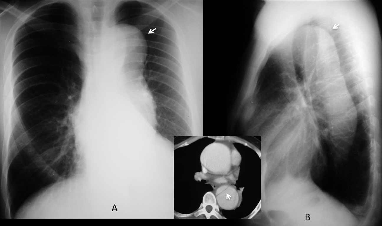
The aortic knob should be examined in every chest radiograph. The most obvious finding is a change in size. The standard size of the aortic knob is about 35 mm, but it varies according to age. It is small in the young and enlarges progressively with aging, as the aorta unfolds. A large aortic knob in the elderly with a prominent ascending and descending aorta is no cause for concern (Fig. 1). However, a large aortic knob and descending aorta with a non-enlarged ascending aorta should raise the possibility of a type B aortic dissection (Fig. 2).
Fig. 1. Normal small aortic knob in a 23-year-old man (A, arrow). 75-year-old man with a large aortic knob (B, arrow) and prominent ascending and descending aorta. This appearance is considered normal for the age.
Fig. 2. Asymptomatic 46-year-old man with a large aortic knob and descending aorta (A and B, arrows). The ascending aorta is not prominent. This appearance is suggestive of Type B dissection, confirmed with enhanced CT (insert, arrow).
A large aortic knob with no other changes suggests a localized aneurysm or aortic dissection, especially if the patient is not elderly (Fig. 3).
Fig. 3. 54-year-old patient with a large aortic knob (A, arrow). Enhanced coronal CT confirms an aneurysm of the aortic arch (B, arrow).
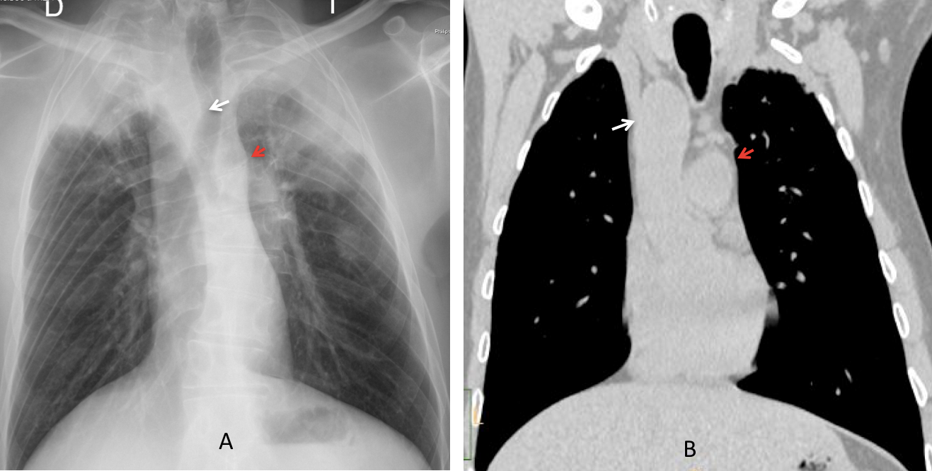
As was mentioned, small aortic knob is common in youth. A small or non-existent left aortic knob in adults is usually because it is on the opposite side (right aortic arch). This congenital variation is easily detected by recognizing the knob at the right mediastinal border and detecting the imprint in the right tracheal wall (Fig. 4). Recognizing these findings can be helpful in selected cases (Fig. 5).
Fig. 4. 27 year-old woman with no obvious left aortic knob, which is located instead on the right side (A, white arrow). Note the imprint in the right tracheal wall (A, red arrow). Angiography confirms the right aortic arch (B, white arrow). The small bump under the right arch represents the azygos vein (A and B, yellow arrows).
Fig. 5. 35 year-old woman with bilateral pneumonia and a possible right mediastinal mass imprinting the trachea (A, white arrow). Unenhanced coronal CT shows that the mass corresponds to a right aortic arch (B, white arrow). The apparent small left aortic arch in the plain film (A, red arrow) is due to the main pulmonary artery, which is elevated because of the patient’s scoliosis (B, red arrow).
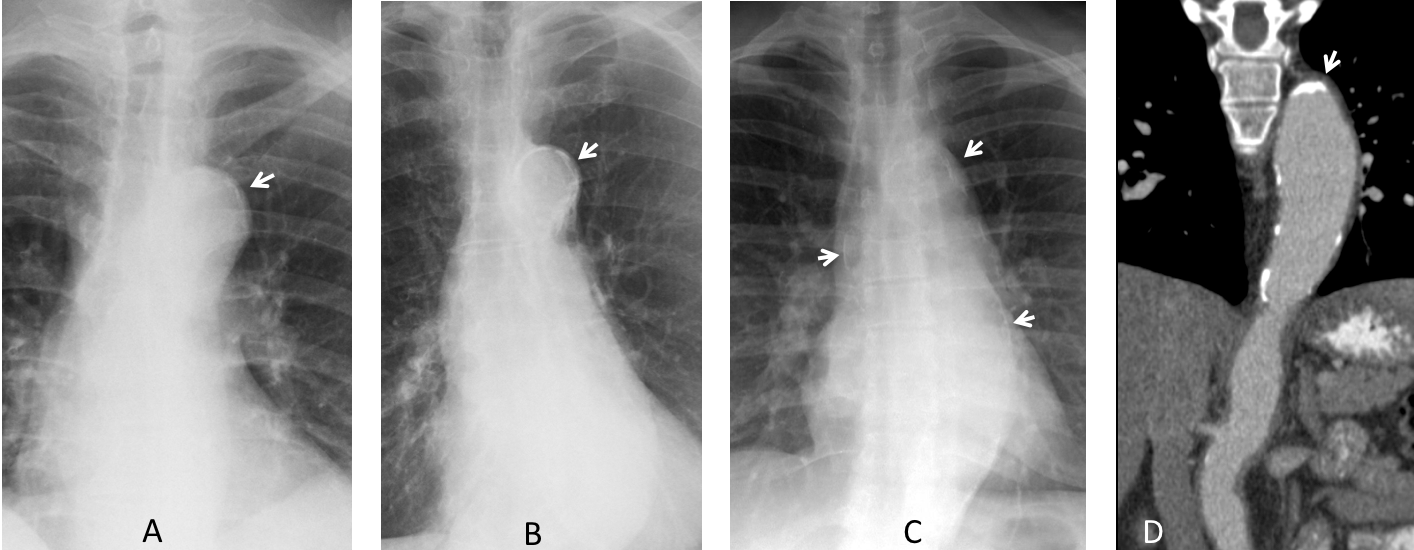
Calcification of the aortic knob is normal in the elderly, but should be considered abnormal in young patients. If conditions that produce hypercalcemia are excluded, the possibility of aortitis should be considered (Fig. 6).
Fig. 6. Two patients aged 65 and 77 years with knob calcification (A and B, arrows), a finding normal for their ages. The third patient is 40-year-old and has calcifications in the aortic knob and ascending and descending aorta (C, arrows). This finding is abnormal and should be investigated. Coronal CT shows aortic calcifications and irregularity (D, arrow). Diagnosis: Takayasu disease
(Case courtesy of Eva Castañer, MD)
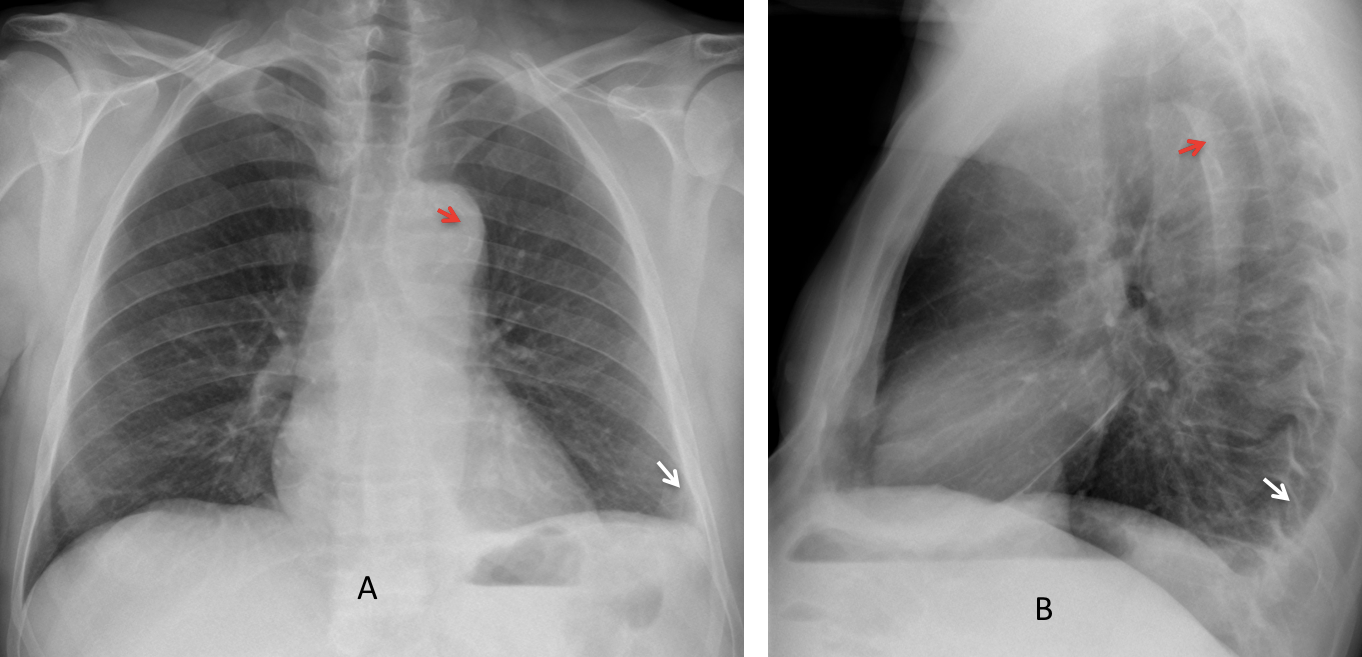
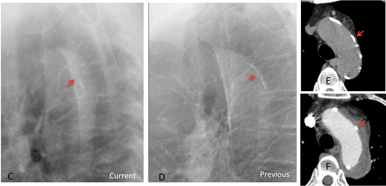
Medial displacement of intimal calcium has been said to be a good sign of aortic dissection. In my experience this sign is unreliable, although it is helpful at times, especially if we have a previous film to compare (Fig. 7).
Fig. 7. 52 year-old man with chest pain. Chest radiographs show a small left pleural effusion (A and B, white arrows). Curvilinear intimal calcium seems to be displaced within the aortic lumen (A and B, red arrows).
Comparison with a previous lateral view confirms displacement of the intimal calcium (C, arrow), which was located in the periphery in the earlier film (D, arrow).
A previous axial CT shows the calcium in the aortic wall (E, arrow). Current enhanced CT depicts the dissection surrounding the aortic lumen with the calcium displaced inward (F, arrow).
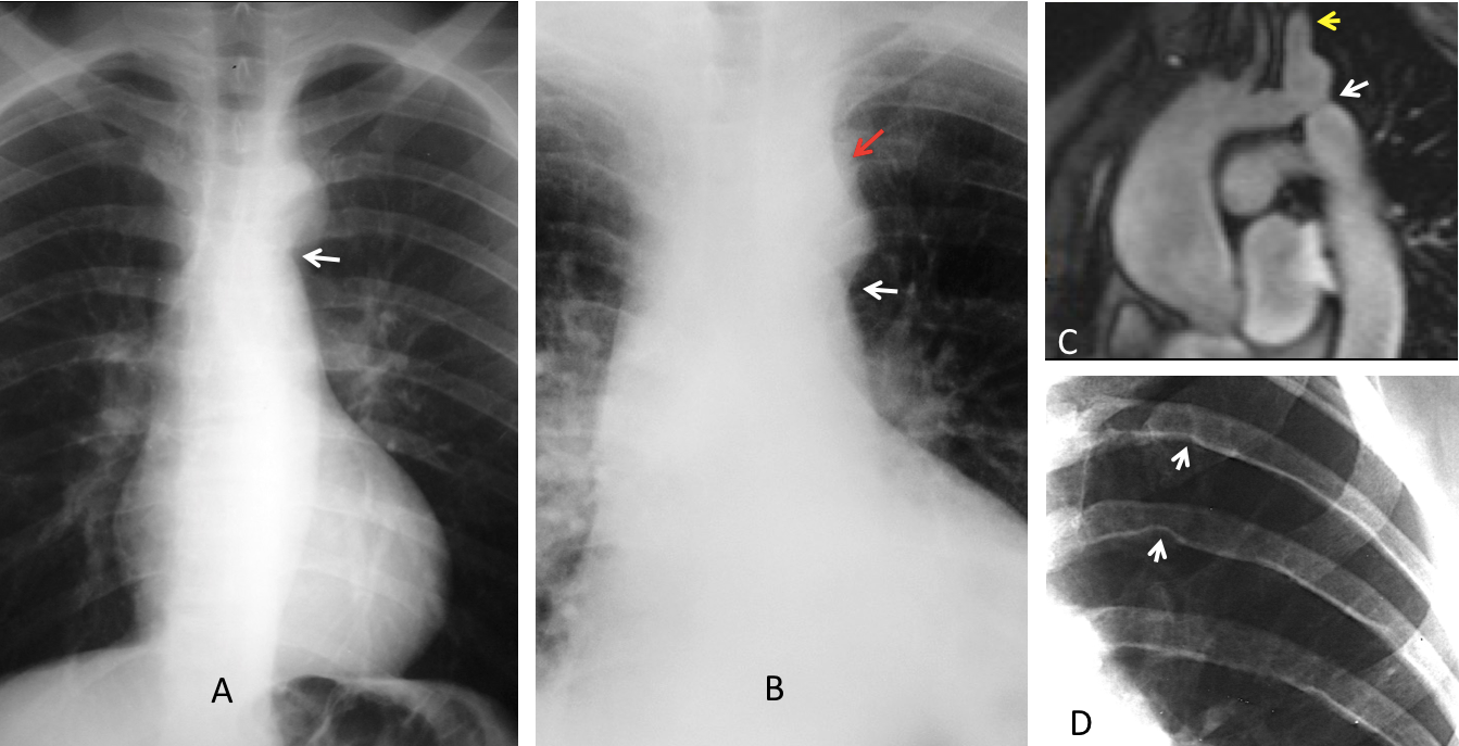
Changes in the contour of the aortic knob may be helpful for diagnosing specific conditions. The best known is the figure of 3 sign, indicative of aortic coarctation. An additional sign is blurring of the upper contour of the knob, due to the dilated left subclavian artery. It is accompanied by rib notching, secondary to collateral circulation (Fig. 8).
Fig. 8. Two cases of aortic coarctation showing the typical figure of 3 sign (A and B, white arrows). The second case also shows obliteration of the upper knob due to the enlarged left subclavian artery (B, red arrow). MRI depicts the area of narrowing (C, white arrow), and the dilated left subclavian artery (C, yellow arrow). Rib notching is evident (D, arrows)
Aortic pseudocoarctation is an uncommon malformation in which there is kinking of the aorta distal to the subclavian artery, without narrowing. The appearance on plain films is similar to that of true coarctation (Fig. 9). There is no significant obstruction and no gradient across the kinking. Patients do not have hypertension or rib notching.
Fig. 9. Pseudocoarctation with figure of 3 knob (A, arrow). Angiogram shows kinking without narrowing (B, arrow).
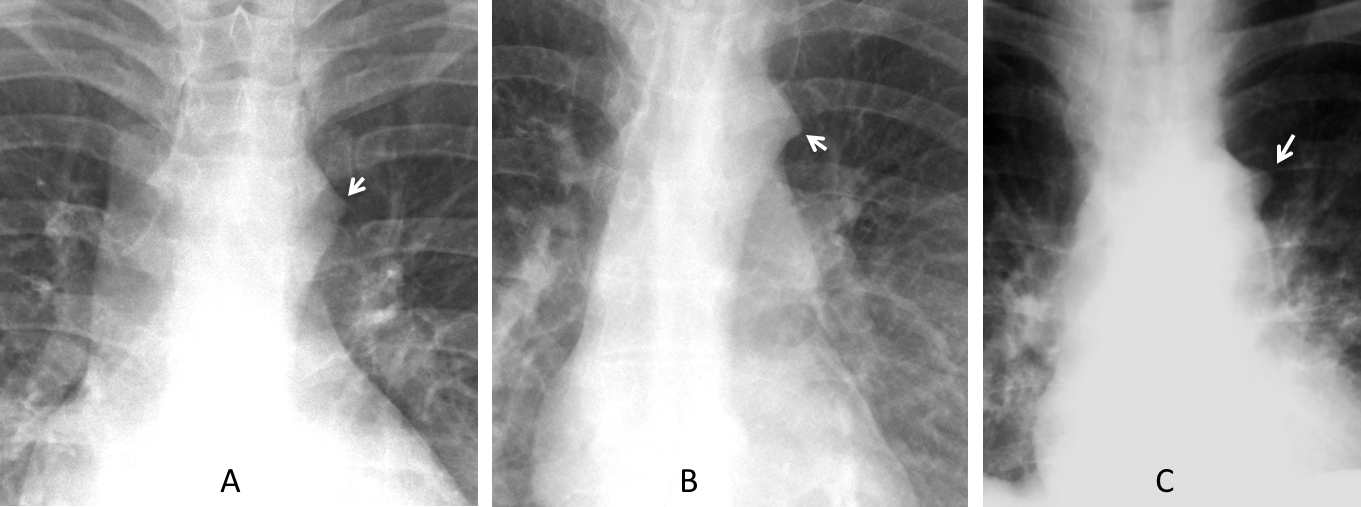
The feature known as aortic nipple is a common change in the contour, visible in up to 10% of PA radiographs. It results from the left superior intercostal vein circling around the aortic knob. A large nipple suggests increased flow in the left hemiazygos system (Fig. 10).
Fig. 10. The first two images show the normal appearance of aortic nipple (A and B, arrows). The third and fourth images show an enlarged aortic nipple (C and D, arrows), which should be investigated.
Unenhanced coronal CT confirms a large aortic nipple (E, white arrow), which corresponds to an enlarged left superior intercostal vein, better seen in the axial CT (F, white arrow). There is enlargement of the azygos vein as well (E and F, red arrows). Diagnosis: congenital absence of inferior vena cava with azygos continuation.
Last but not least, a double contour of the aortic knob should raise the possibility of a superimposed mediastinal mass, either in front of or behind the aortic knob (Fig. 11).
Fig. 11. 64-year-old woman with back pain, who had a well-differentiated liposarcoma removed from her right thigh seven years earlier. PA radiograph shows a double contour of the aortic knob (A, arrows). It does not obliterate the aortic contour, indicating a mediastinal mass in front of or behind the aorta. Coronal CT demonstrates a posterior mediastinal soft tissue mass (B, arrow). MRI shows the mass with involvement of the adjacent vertebra (C, arrow). Diagnosis: vertebral metastases from liposarcoma.
Follow Dr. Pepe’s advice:
1. An enlarged aortic knob in the young adult should be investigated.
2. The most common cause of small or absent left aortic knob is a right aortic arch.
3. Calcification of the knob in the young adult should be investigated.
4. Most common changes in the contour are aortic coarctation and aortic nipple.









Broken ribs on the right side.
Good morning! Fracture of the 5 right costal arc, signs of médium sternotomy.
There is an elongated aorta. Gynecomastia.
There are some nodular opacities proyected over both inferior hemithorax perhaps because of skin lesions (in the lateral view there is a round cutaneal image next to the sternotomy) or pleural calcifications.
There is an aereal image in anterior mediastinum…
Hello. There is a fracture of the rt 6th rib laterally. I see prominence of the aortic arch and left pulmonary artery, and a small nodular opacity immediately above the left hilum. I also think that two inferior fragments of the sternal suture are located too deep.
What do you think about the nodular opacity?
The contours are too rounded to be a vessel. I believe it could be a nodule in the aortopulmonary window, and I think it is visible on the profile image overlying the trachea. I would investigate further with CT. After a second look, I would also say that there is thickening of the rt paratracheal line.
Multiple right rib fractures
Ectopic gas, in subxyphoid position
I’d like to compare w/ previum films, to check the sternotomy “wires”, because they’re broken
I’m answering at a phone, cannot define better if there is or isn’t any pneumothorax.
…fratture costali a dx….rottura dei fili della sternotomia mediana, inferiormente e pneumomediastino….sospetto di doppio contorno dell’arco aortico , come da dissezione……Auguroni PROF. per le festività , per il prestigioso incarico e per il tuo Barca….per me continua la sofferenza per il mio Bari…..ti prego consultare il sito di pediatric radiology VCU e vedere i vincitori per il 2017….
A fracture is seen involving the angle of the 6th right rib,sternotomy wire, Prominence of both breast nipple at the 5th anterior ribs, somewhat patchy opacity at the right lower lung zone, and some calcification on the wall of the descending aorta.
The answer is: aneurysm of aortic arch. Congratulations to Mauro, who discovered the abnormality and placed it in the lateral view.