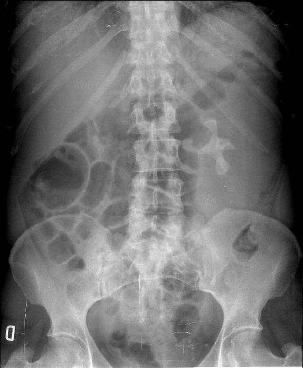
This week’s case is a 51 year-old woman with a history of chronic urinary tract infection. She consulted for left lumbar pain and intermittent mild fever of several weeks’ duration.
1. Staghorn calculus with pyonephrosis
2. Staghorn calculus with hydronephrosis
3. Staghorn calculus and renal tumour
4. None of the above
Findings: The abdominal film shows an enlarged left kidney (
green arrows) with a large staghorn calculus occupying the calyceal-pelvic area (
blue arrow).
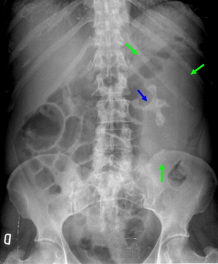
Fig. 2
Axial (Fig. 3) and coronal (Fig. 4) contrast-enhanced CT images confirm an enlarged, non-functioning left kidney with multiple round low-attenuation areas (Fig. 4, red arrows) replacing the renal parenchyma, and a staghorn calculus (blue arrow). Marked perinephric inflammatory changes are also evident, with thickening of the renal fascia and abdominal wall (green arrows). These findings are highly suggestive of xanthogranulomatous pyelonephritis.
Final diagnosis: Xanthogranulomatous Pyelonephritis
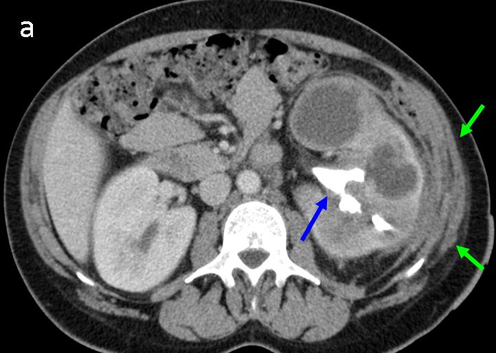
Fig. 3

Fig. 4
Xanthogranulomatous pyelonephritis (XGP) can be suspected in abdominal films when a diffusely enlarged kidney associated with a staghorn calculus is seen in a patient with a history of chronic urinary tract infection, consulting for malaise and low-grade fever.
CT is the imaging technique of choice for the final diagnosis of XGP because it conclusively confirms the enlarged, poorly functioning or non-functioning kidney and staghorn calculus, and demonstrates the typically multiple, round, low attenuation areas, which represent dilated calyces and pus-filled spaces replacing the renal parenchyma.
CT also clearly shows the severity and extension of inflammatory involvement in adjacent organs and affected tissues, very common features in this inflammatory renal process. Perirenal abscess and fistulas to contiguous organs and soft tissues adjacent to the affected kidney are very common in this disease.
CT is determinant in cases in which abdominal films show only the staghorn calculus, without obvious enlargement of the kidney (Fig. 5, A-C)
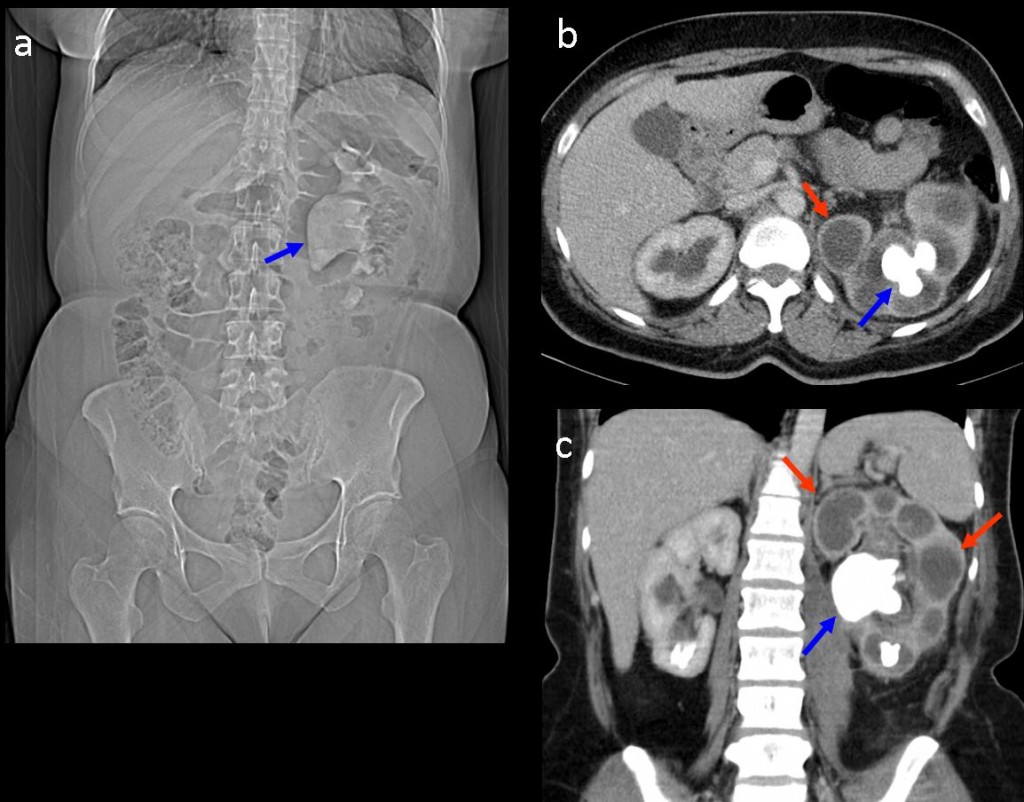
Fig. 5
In this case, the plain film (Fig. 5a) only shows a staghorn calculus (blue arrow) in an apparently normal sized left kidney. Enhanced CT (Fig. 5b-c) shows typical findings of XGP: multiple, round, hypodense, low attenuation areas (red arrows) in an enlarged non-functioning left kidney and the obstructive staghorn calculus (blue arrows).
Staghorn calculus can cause chronic urinary tract obstruction with severe hydronephrosis and renal parenchyma atrophy, but without inflammatory disease. In this patient, a chronic obstructive left staghorn lithiasis (Fig. 6 blue arrows in a-c) has caused severe renal atrophy with hydronephrosis (red arrows in 6b-c)
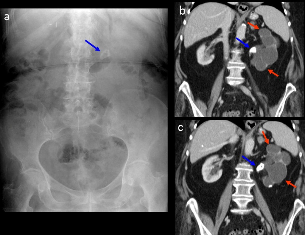
Fig. 6
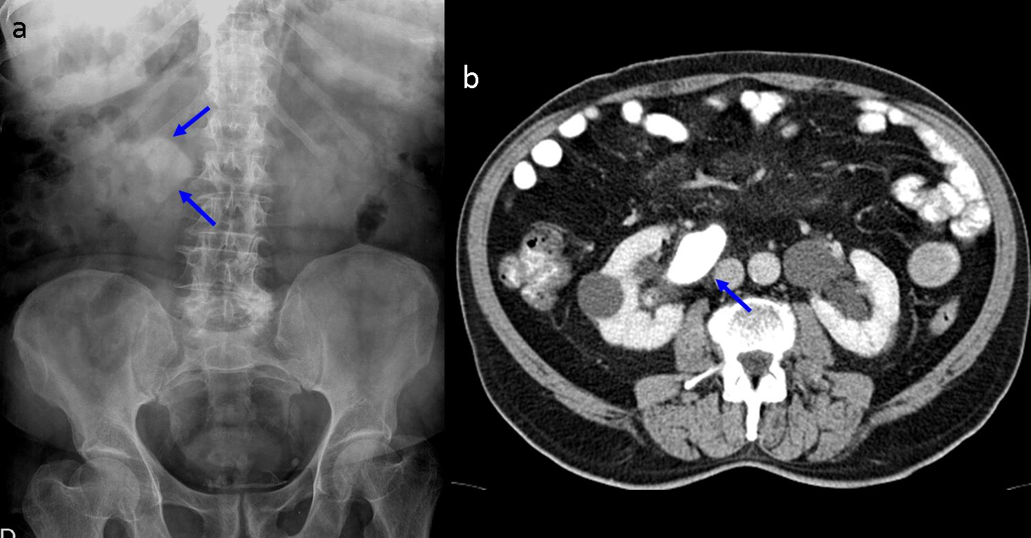
Staghorn calculus can be present without urinary tract obstruction or inflammatory renal disease. In this patient with a right kidney staghorn calculus detected on the abdominal plain film (Fig. 7 blue arrows), the contrast-enhanced CT confirms the lithiasis (Fig. 7b blue arrow), but there is no urinary tract obstruction or inflammatory lesions of the kidney (b).
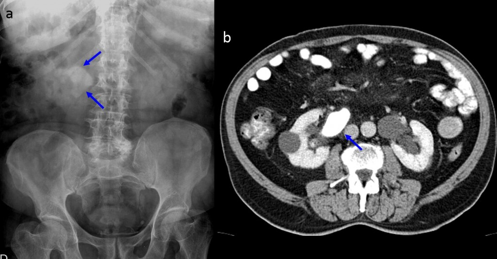
Fig. 7

Follow Dr. Pepe’s advice
Follow Dr. Pepe’s advice:
- In a patient with persistent low-grade fever in whom the abdominal film shows a staghorn calculus and enlarged kidney, XGP should be suspected
- CT is diagnostic by showing a non-functioning kidney with round, low-attenuation areas and perirenal involvement
- CT also differentiates XGP from severe hydronephrosis and non-obstructive staghorn calculus
Suggested reading: Xanthogranulomatous Pyelonephritis. RadioGraphics 11(3):485-498, 1991
Case prepared by Julio Martin MD