Dr. Pepe’s Diploma Casebook: Case 18 – SOLVED!

Dear Friends,
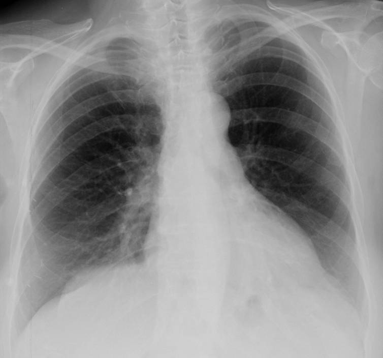
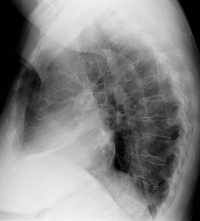
My second case of 2013 relates to a 49-year-old man with shortness of breath. PA and lateral radiographs are shown.
Diagnosis:
1. McLeod syndrome
2. Pulmonary embolism
3. Pneumothorax
4. None of the above
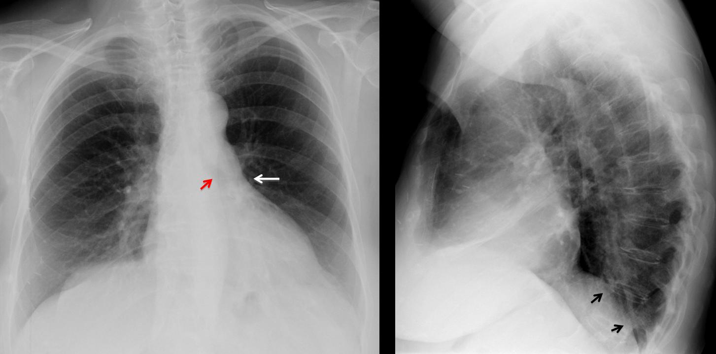
Fig. 1
Findings: PA film shows increased lucency of the left lung. The left hilum appears small because it is descended and hidden behind the cardiac silhouette (arrow). The left bronchus is vertically oriented (red arrow). On the lateral view, there is blurring of the left hemidiaphragm (arrows). These signs are characteristic of LLL collapse with increased lucency of left lung due to compensatory expansion of LUL.
Coronal MIP reconstruction confirms the descent of the left hilum and the vertical orientation of the main left bronchus (arrow). No endobronchial lesion is visible. Coronal CT shows loss of volume of LLL with medial displacement of major fissure (arrow) and bronchiectasis (red arrows). There is compensatory elevation of the left hemidiaphragm.
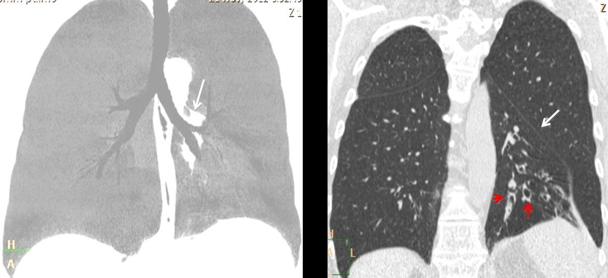
Fig. 2
Final diagnosis: LLL collapse, secondary to bronchiectasis.
The typical appearance of LLL collapse is that of a triangular opacity seen through the heart in the PA view and as posterior lower triangular opacity in lateral view. Additional signs are: descended left hilum, which appears small because it is hidden behind the heart, and increased lucency of the left lung secondary to compensatory expansion of LUL. These changes are described in Fig. 3, below.
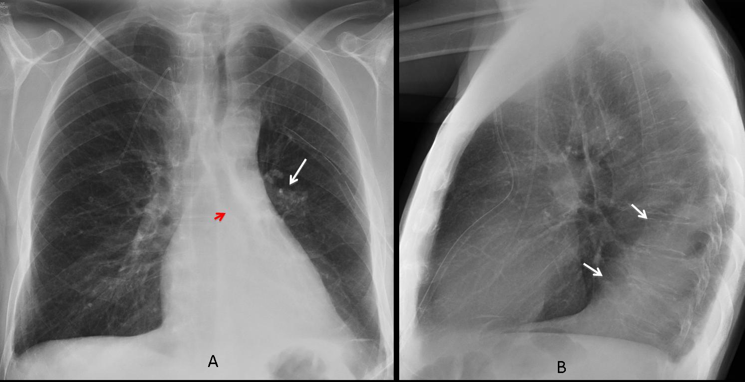
Fig. 3
Fig. 3 A & B (above): transient LLL collapse secondary to mucous plug. PA chest shows the hyperlucent LUL lobe, downward displacement of left hilum (arrow) with vertically–oriented left bronchus (red arrow). Increased retrocardiac triangular opacity, better seen in the lateral view (arrows).
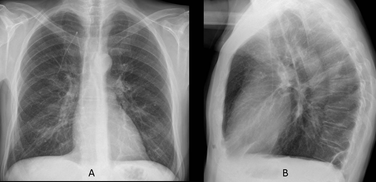
Fig. 4
Fig. 4 A & B (above): radiographs taken six days later, after removal of the mucous plug, show that the left hilum has returned to the normal location and the triangular opacity of LLL collapse has disappeared, as well as the increased lucency of LUL.
In extreme cases of LLL collapse, the increased lucency of the expanded LUL may predominate, giving the deceiving appearance of a hyperlucent left lung. The collapsed LLL lobe is barely visible in the PA view. On the lateral view only blurring of the left hemidiaphragm is seen. The clue to a correct diagnosis lies in correctly identifying the downward displacement of the left hilum (Figs 5-7).
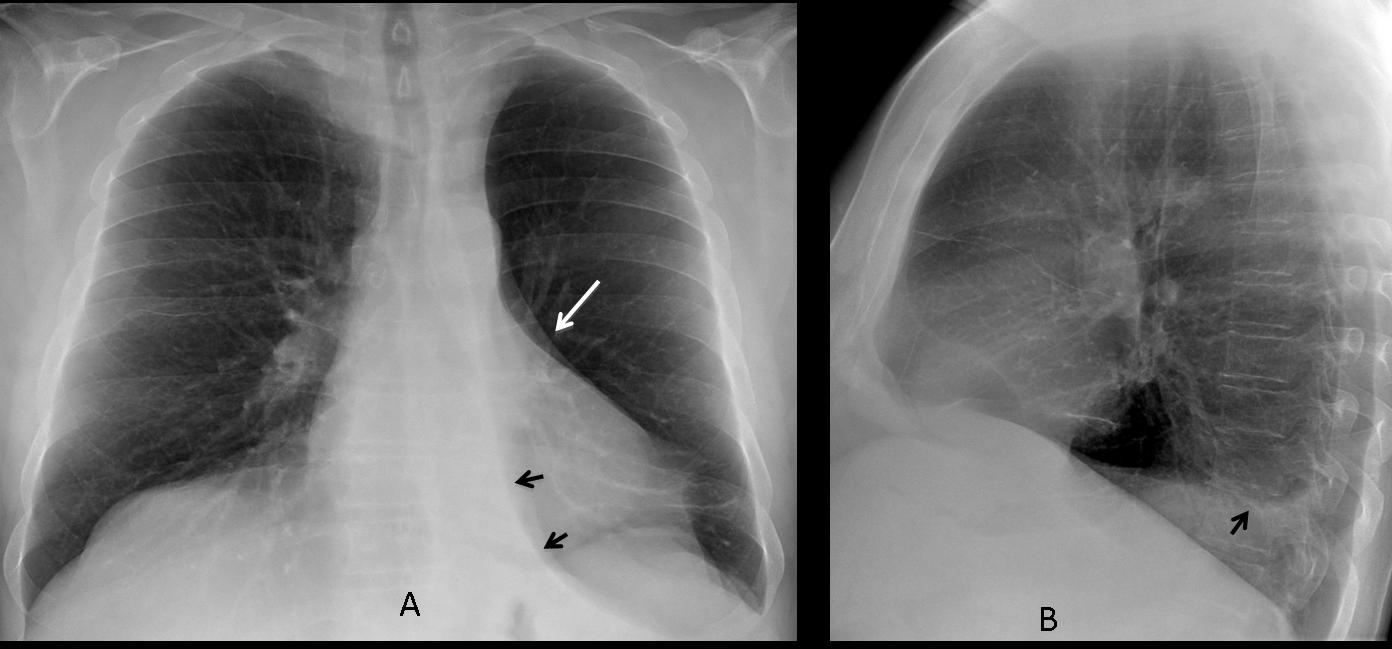
Fig. 5
Fig. 5 A & B (above): pre-op film in an asymptomatic 68-year-old man with marked LLL collapse simulating hyperlucent lung. The collapsed lobe is seen as a small paramediastinal triangular opacity (arrows) and slight blurring of hemidiaphragm in the lateral view (arrow). The clue to the diagnosis is the marked downward displacement of the left hilum (white arrow).
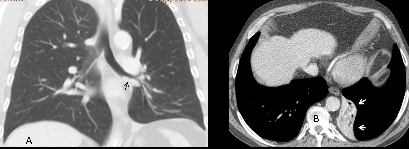
Fig. 6
Fig. 6 A & B (above): coronal CT confirms the downward displacement of hilum and an endobronchial lesion in the LLL wall (C, arrow). The axial view shows the marked collapse of LLL (arrows). Final diagnosis: unsuspected squamous-cell carcinoma of LLL bronchus.
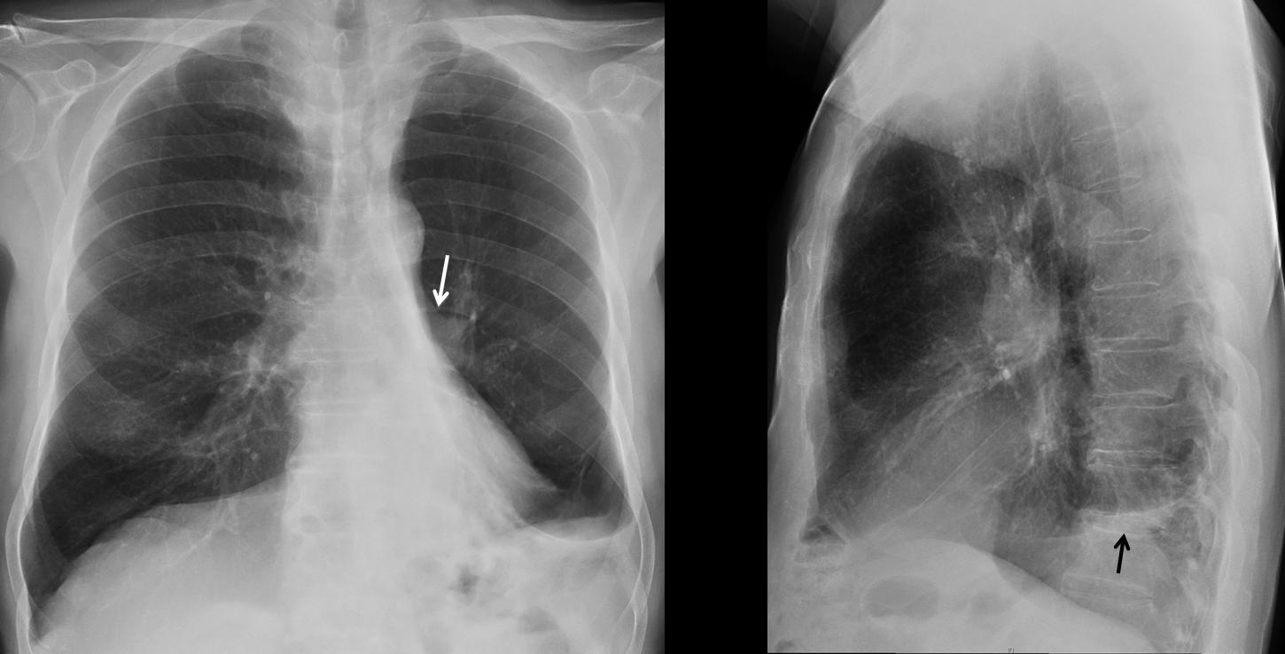
Fig. 7
Fig 7 (above): bronchiectasis with marked LLL collapse. Hyperlucent lung with marked downward displacement of left hilum (arrow) and haziness of hemidiaphragm (arrows). Note the similarity to the previous case.
Although bronchiectasis alone may cause collapse of LLL lobe CT should be done to rule out an endobronchial lesion.
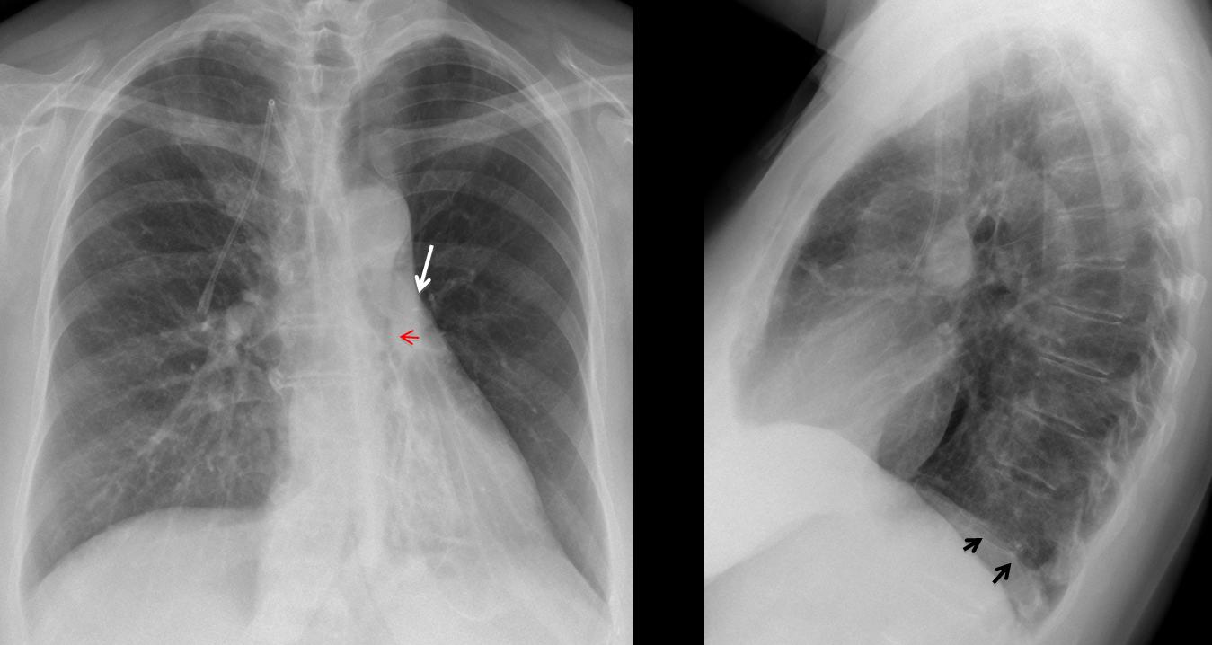
Fig. 8
Fig. 8 (above): 56-year-old female with extreme collapse of LLL attributed to bronchiectasis. Note the marked hyperlucency of the LUL and the downward displacement of the left hilum (arrow). There is a suggestion of an endobronchial lesion in the PA view (red arrow). There is minimal blurring of hemidiaphragm in the lateral view (arrows).
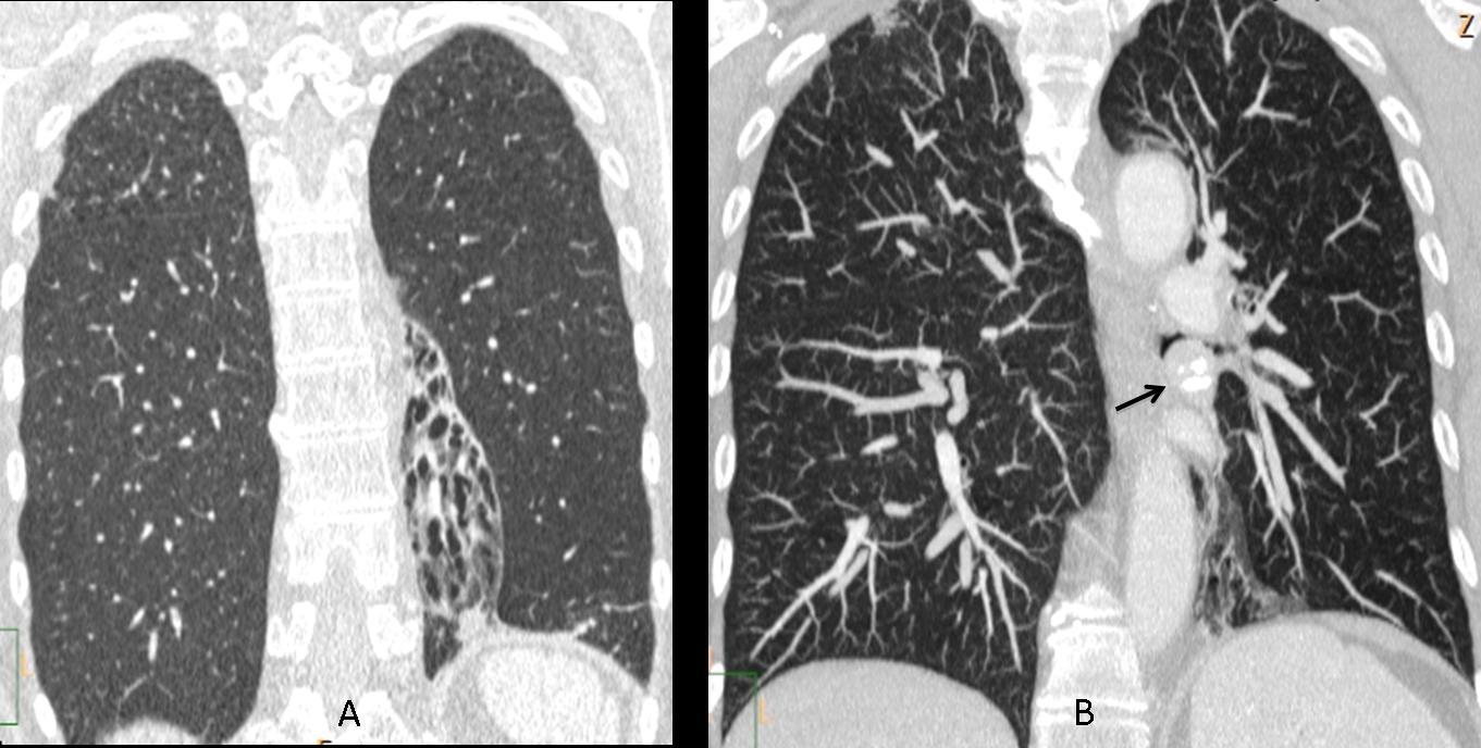
Fig. 9
Fig. 4 A & B (above): coronal CT shows marked LLL volume loss with bronchiectasis secondary to a rounded endobronchial lesion with popcorn calcification (B, arrow). Final diagnosis: endobronchial neuroendocrine tumour.
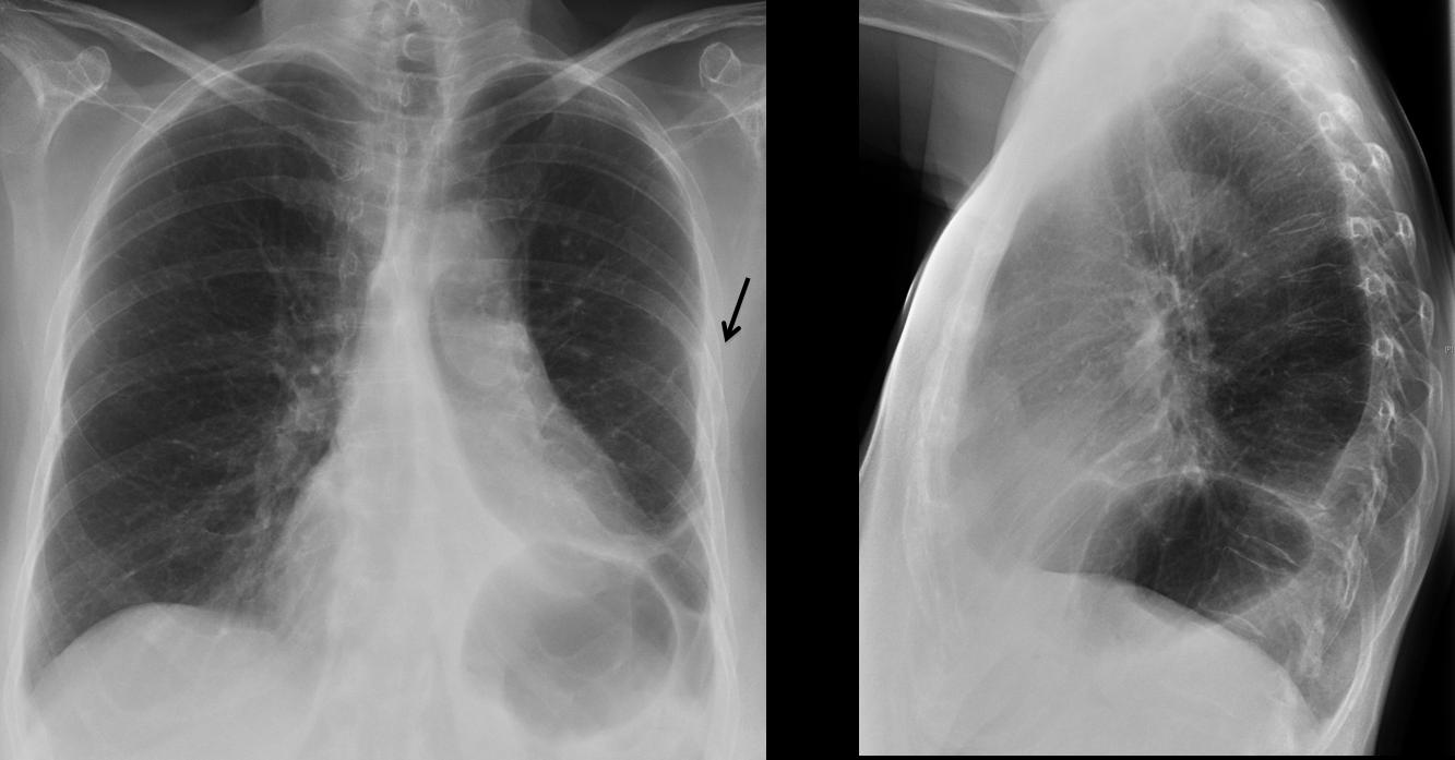
Fig. 10
Fig 10 (above): surgical resection of LLL may simulate extreme LLL collapse, as in the case above. Note the hidden left hilum, elevation of left hemidiaphragm and triangular paramediastinic opacity. The proximity of the 5th and 6th ribs are a clue to suspect previous surgery (arrow).

Follow Dr. Pepe’s advice
Follow Dr. Pepe’s advice:
- In extreme cases of LLL collapse the compensatory expansion of LUL may predominate, suggesting a hyperlucent lung
- Downward displacement of left hilum, verticalization of left bronchus and haziness of the diaphragmatic contour on the lateral view should suggest the correct diagnosis
- In all cases, CT should be done to exclude an endobronchial lesion
Recommended reading: Unusual pattern of left lower lobe atelectasis. Radiology 141: 331-333, 1981
Case prepared by Dr. Pepe


