This week, we have an oncologic patient with liver nodule detected on US examination. Below are the images from an MRI examination.
1. Liver hemangioma
2. Hepatocellular carcinoma (HCC)
3. Liver metastasis
4. Focal nodular hyperplasia (FNH)
Findings: T2-W image (a) depicts a slightly hyperintense liver nodule in segment VII that is hypointense in the T1-W sequence (b) (white arrows). The nodule shows considerable hypervascularity on dynamic arterial imaging (c), and remains hyperenhanced in the venous phase (d) (yellow arrows).
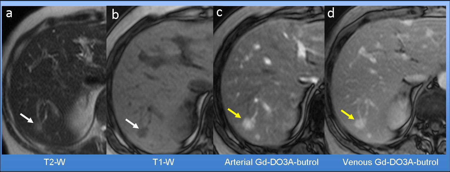
Fig. 1
MR findings exclude the diagnosis of liver hemangioma (option 1) because hemangiomas are strongly hyperintense on T2-W images (Fig. 2a, arrow).
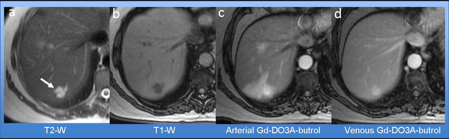
Fig. 2
Hepatocellular carcinoma (option 2) can be eliminated because the lesion usually shows washout in the venous phase (Fig. 3h, arrow).
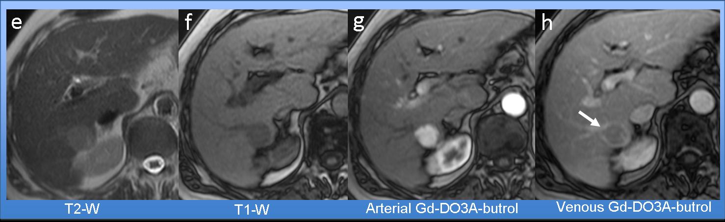
Fig. 3
The remaining diagnoses are liver metastasis (option 3) and FNH (option 4). With the use of gadoxetic acid disodium, non-hepatocellular lesions, such as liver metastasis, can be easily differentiated from hepatocellular lesions, such as FNH. Fig. 4 shows images following gadoxetic acid disodium injection. The findings are similar to those obtained with conventional contrast agent except for the late liver-specific phase (d, green arrow), in which the nodule is clearly hypointense because of the absence of hepatocytes. The red arrow indicates contrast excretion from the biliary tree.
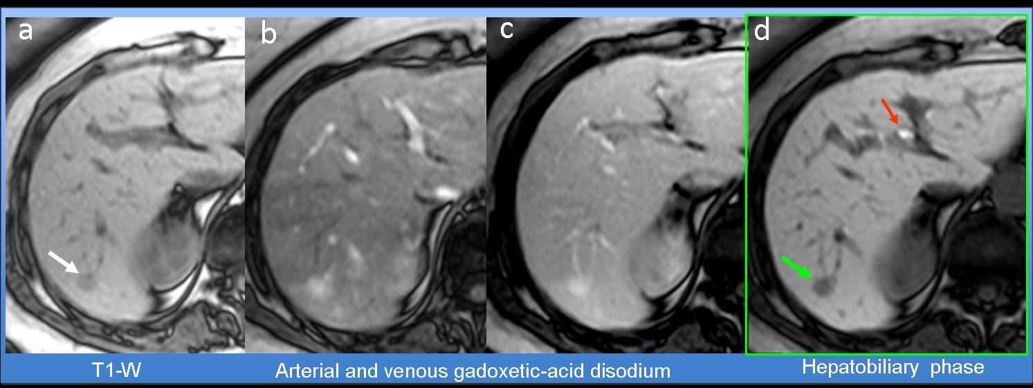
Fig. 4
Final diagnosis: Hypervascular liver metastasis from medullary thyroid carcinoma
In studies using gadoxetic acid contrast agent, hepatocellular lesions such as FNH are hyperintense in the hepatobiliary phase (Fig. 5h, green arrow) confirming the hepatocellular nature of the lesion.

Fig. 5
Gadoxetic acid disodium is a relatively recent gadolinium-based contrast agent for T1-W MR imaging of the liver that accumulates in functioning hepatocytes during the hepatobiliary phase (10-15 min after administration). This agent combines the properties of conventional extracellular fluid contrast agents and hepatobiliary agents, enabling dynamic perfusion imaging and evaluation of delayed hepatocyte uptake.
Non-hepatocyte-containing lesions, such as metastases, are invariably hypointense on images obtained in the hepatobiliary phase, whereas benign hepatocellular lesions or tumour-like lesions, such as focal nodular hyperplasia, display hepatocyte-selective uptake and are isointense, or more commonly hyperintense, relative to the liver parenchyma, confirming the hepatocellular nature of the lesion.
In cross-sectional CT or dynamic MR examinations using conventional extracellular contrast agents, hypervascular liver metastasis from some primary neoplasms, such as kidney, thyroid or neuroendocrine tumours, can have features very similar to those of other benign lesions, like FNH. In these cases, gadoxetic acid-enhanced MR imaging during the late hepatobiliary phase helps to characterise the lesion and resolve doubts, as hepatocyte-containing nodules will show contrast uptake, whereas non-hepatocellular lesions will not.

Follow Dr. Pepe’s advice
Follow Dr. Pepe’s advice:
- Hypervascular liver metastasis can be difficult to differentiate from benign lesions like FNH on dynamic CT or MR examination using conventional extracellular contrast agents.
- Delayed hepatobiliary phase MR imaging using liver-specific gadoxetic acid disodium can help to achieve the correct diagnosis in these cases.
- Lesions showing contrast uptake similar to or higher than background liver parenchyma in the hepatobiliary phase are hepatocellular lesions, whereas metastases are always hypointense relative to liver parenchyma.
Recommended reading: MR Imaging in Patients with Suspected liver Metastases: Value of Liver-specific Contrast Agent Gd-EOB-DTPA . Magn Reson Med Sci, Vol. 6, Nº. 1, pp 43-52, 2007
Case prepared by Julio Martin MD