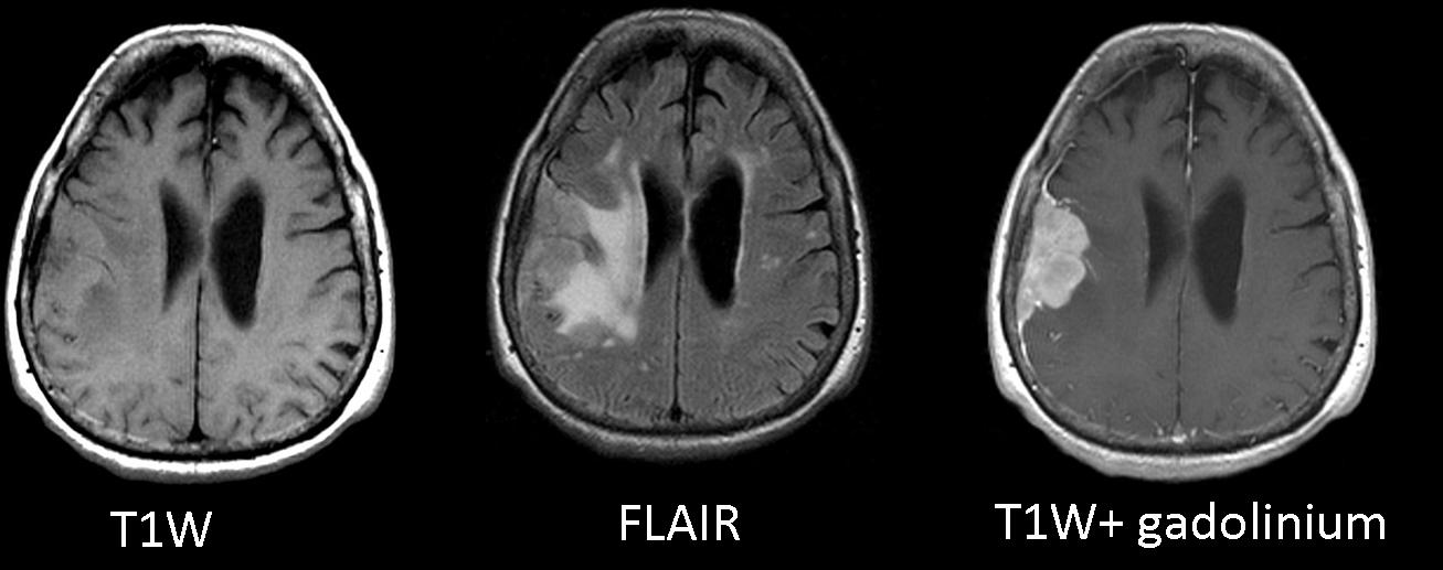
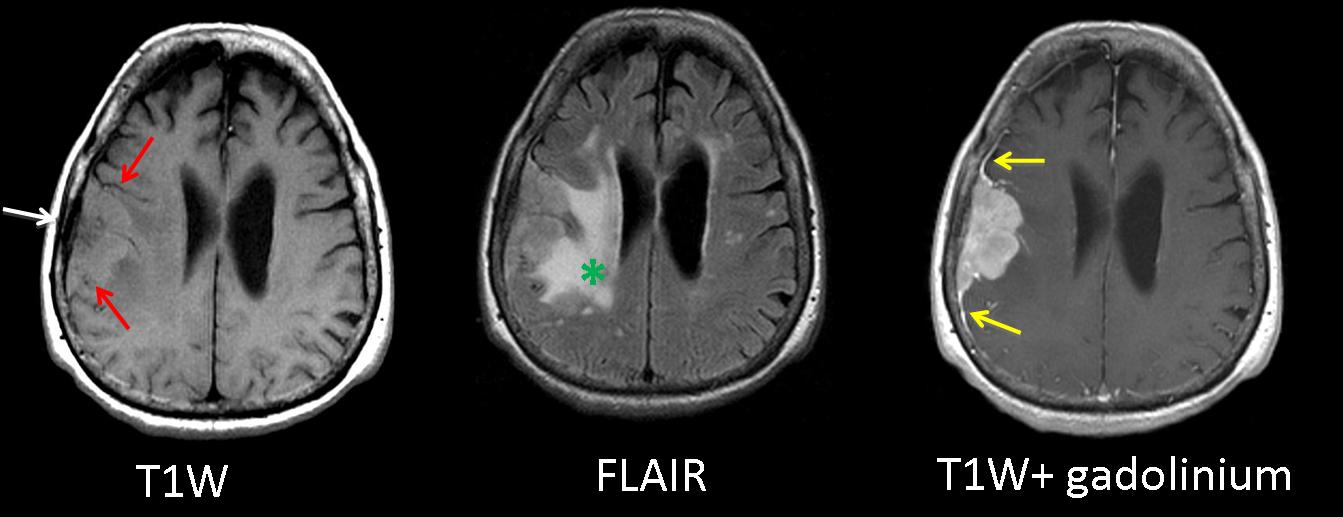
Showing MRI images of an 82-year-old woman with partial motor seizures afecting her face.
T1W MR image shows a right dural-based, extra-axial, frontoparietal mass (
red arrows) and intraparenchymal edema (
green asterisk) on FLAIR sequence. The mass enhances markedly on T1W gadolinium-enhanced imaging and shows a dural tail (
yellow arrows) There is thickening of the inner table wall (white arrow).
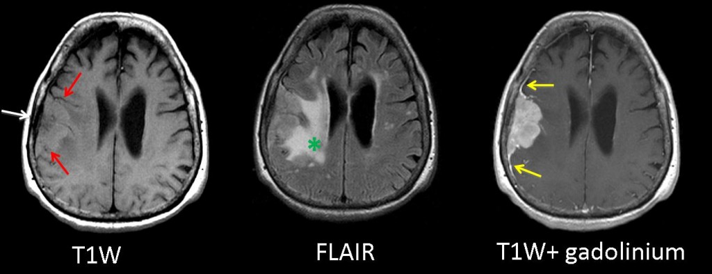
Fig. 1
Conclusion: Extra-axial homogenous enhancing mass with associated thickening of the inner table wall, medial displacement of the cortex, and dural enhancement
Final diagnosis: WHO Grade II Meningioma
The differential diagnosis of extra-axial cranial masses should include meningiomas, metastatic tumors, multiple myeloma, lymphoma, chloroma, Rosai-Dorfman disease, and sarcoidosis.
Meningiomas are the most common nonglial primary tumors of the central nervous system and the most common extra-axial neoplasms, accounting for approximately 15% of all intracranial tumors.
The typical meningioma is a homogeneous, hemispheric, markedly enhancing extra-axial mass. The dural tail sign on gadolinium MR images refers to a thickening of the enhanced dura that simulates a tail extending from the mass. It appears in 60-72% of meningiomas, but it can also be seen in other dural-based masses (lymphoma, metastasis, chloroma, sarcoidosis, plasmacytoma, neurinoma). Meningiomas may be associated with hyperostosis.
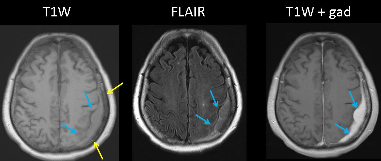
Multiple Myeloma
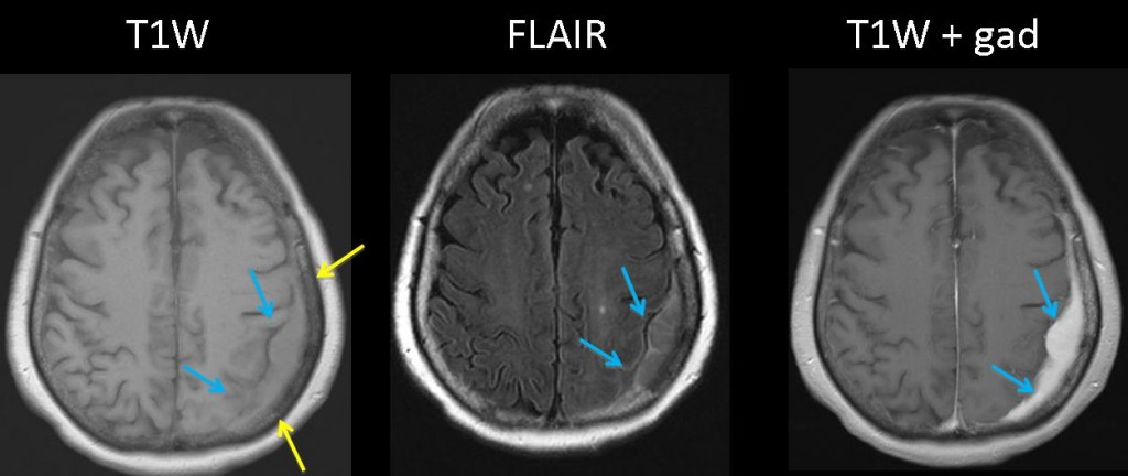
Fig. 2
71-year-old woman with multiple myeloma who presents with ataxia.
MR images show a left extra-axial mass with intermediate signal on T1W and FLAIR images, and homogeneous increased signal on gadolinium-enhanced images (blue arrows). T1W images show focal low signal intensity of the infiltrated bone marrow adjacent to the mass (yellow arrows)
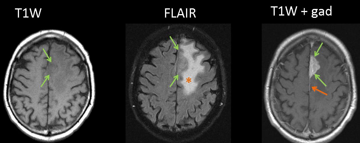
Dural metastasis
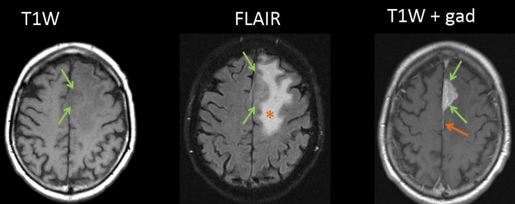
Fig. 3
72-year-old man diagnosed of squamous cell lung carcinoma with right hemiparesis.
MR images show a left frontal extra-axial, dural-based mass (green arrows), with intermediate signal on T1W and FLAIR sequences, homogeneous enhancement on gadolinium-enhanced images, and linear dural enhancement (orange arrows). Adjacent intraparenchymal edema is better demonstrated on FLAIR images as an area of increased signal (asterisk).

Follow Dr. Pepe’s advice
Follow Dr. Pepe’s advice:
- Meningiomas are the most common extra-axial neoplasms.
- Typical meningiomas present as markedly enhancing extra-axial masses. The dural tail sign, although non-specific, is found in 60% of them.
- Differential diagnosis should include metastatic tumors, multiple myeloma, lymphoma, chloroma, Rosai-Dorfman disease, and sarcoidosis.
Recommended reading:
1-Patterns of Contrast Enhancement in the Brain and Meninges. RadioGraphics 2007; 27:525–551.
2-Magnetic resonance imaging of meningiomas.Semin Ultrasound CT MR. 1992 Jun;13(3):154-69.
Case presented by Laura Oleaga, MD